

Boundary-Preserved Deep Denoising of Stochastic Resonance Enhanced Multiphoton Images
- PMID: 36304843
- PMCID: PMC9592049
- DOI: 10.1109/JTEHM.2022.3206488
Boundary-Preserved Deep Denoising of Stochastic Resonance Enhanced Multiphoton Images
Abstract
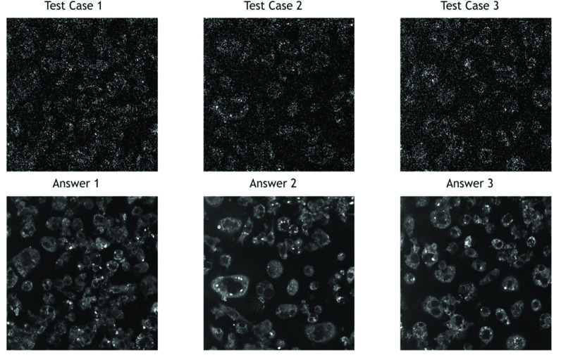
Objective: With the rapid growth of high-speed deep-tissue imaging in biomedical research, there is an urgent need to develop a robust and effective denoising method to retain morphological features for further texture analysis and segmentation. Conventional denoising filters and models can easily suppress the perturbative noise in high-contrast images; however, for low photon budget multiphoton images, a high detector gain will not only boost the signals but also bring significant background noise. In such a stochastic resonance imaging regime, subthreshold signals may be detectable with the help of noise, meaning that a denoising filter capable of removing noise without sacrificing important cellular features, such as cell boundaries, is desirable.
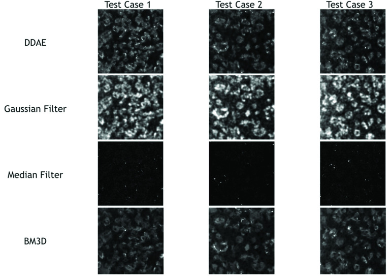
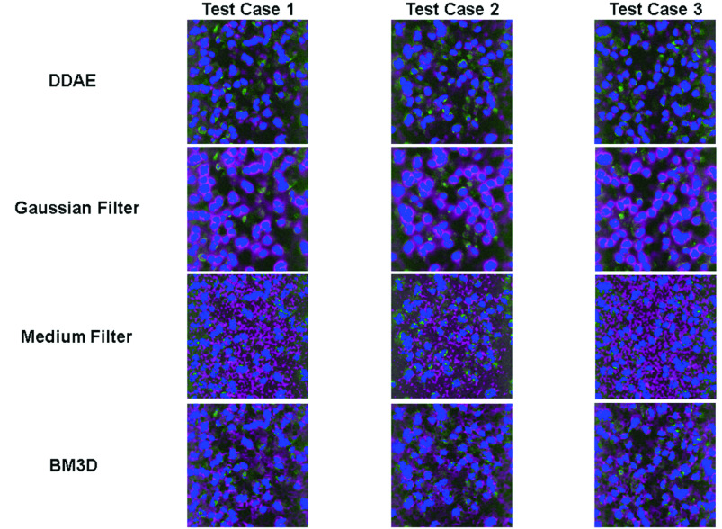
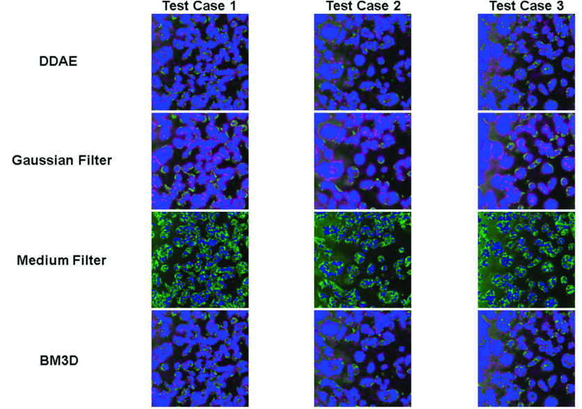
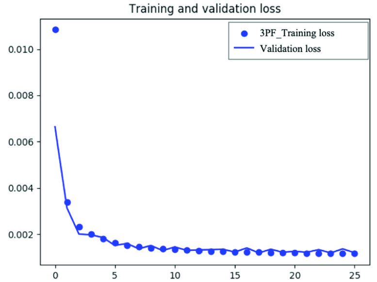
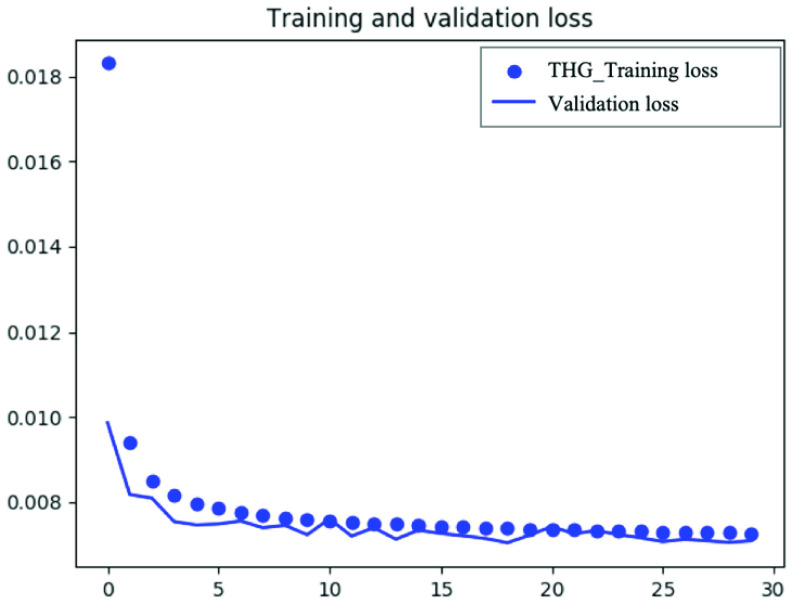
Method: We propose a convolutional neural network-based denoising autoencoder method - a fully convolutional deep denoising autoencoder (DDAE) - to improve the quality of three-photon fluorescence (3PF) and third-harmonic generation (THG) microscopy images.
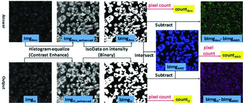
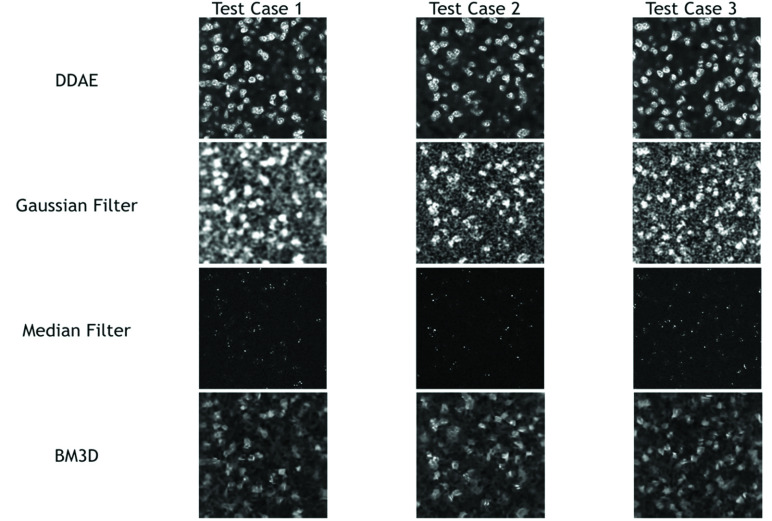
Results: The average of 200 acquired images of a given location served as the low-noise answer for the DDAE training. Compared with other conventional denoising methods, our DDAE model shows a better signal-to-noise ratio (28.86 and 21.66 for 3PF and THG, respectively), structural similarity (0.89 and 0.70 for 3PF and THG, respectively), and preservation of the nuclear or cellular boundaries (F1-score of 0.662 and 0.736 for 3PF and THG, respectively). It shows that DDAE is a better trade-off approach between structural similarity and preserving signal regions.
Conclusions: The results of this study validate the effectiveness of the DDAE system in boundary-preserved image denoising.
Clinical impact: The proposed deep denoising system can enhance the quality of microscopic images and effectively support clinical evaluation and assessment.
Keywords: Third harmonic generation; deep denoising autoencoder; three-photon fluorescence.
Figures
Similar articles
-
Performance of a deep learning-based CT image denoising method: Generalizability over dose, reconstruction kernel, and slice thickness.Med Phys. 2022 Feb;49(2):836-853. doi: 10.1002/mp.15430. Epub 2022 Jan 19. Med Phys. 2022. PMID: 34954845
-
Self-Supervised Image Denoising of Third Harmonic Generation Microscopic Images of Human Glioma Tissue by Transformer-Based Blind Spot (TBS) Network.IEEE J Biomed Health Inform. 2024 Aug;28(8):4688-4700. doi: 10.1109/JBHI.2024.3405562. Epub 2024 Aug 6. IEEE J Biomed Health Inform. 2024. PMID: 38801682
-
Denoising of three-dimensional fast spin echo magnetic resonance images of knee joints using spatial-variant noise-relevant residual learning of convolution neural network.Comput Biol Med. 2022 Dec;151(Pt A):106295. doi: 10.1016/j.compbiomed.2022.106295. Epub 2022 Nov 9. Comput Biol Med. 2022. PMID: 36423533
-
Incorporation of residual attention modules into two neural networks for low-dose CT denoising.Med Phys. 2021 Jun;48(6):2973-2990. doi: 10.1002/mp.14856. Epub 2021 Apr 23. Med Phys. 2021. PMID: 33890681
-
Deep learning on image denoising: An overview.Neural Netw. 2020 Nov;131:251-275. doi: 10.1016/j.neunet.2020.07.025. Epub 2020 Aug 6. Neural Netw. 2020. PMID: 32829002 Review.
Cited by
-
Label-free multimodal imaging with simultaneous two-photon and three-photon microscopy and kernel-based nonlinear scaling denoising.Biomed Opt Express. 2023 Dec 6;15(1):114-130. doi: 10.1364/BOE.504550. eCollection 2024 Jan 1. Biomed Opt Express. 2023. PMID: 38223188 Free PMC article.
References
-
- Denk W., Strickler J. H., and Webb W. W., “Two-photon laser scanning fluorescence microscopy,” Science, vol. 248, no. 4951, pp. 73–76, Apr. 1990. - PubMed
-
- Frigault M. M., Lacoste J., Swift J. L., and Brown C. M., “Live-cell microscopy—Tips and tools,” J. Cell Sci., vol. 122, no. 6, pp. 753–767, Mar. 15, 2009. - PubMed
Publication types
MeSH terms
LinkOut - more resources
Full Text Sources
Medical
