Biocompatible and antibacterial Flammulina velutipes-based natural hybrid cryogel to treat noncompressible hemorrhages and skin defects
- PMID: 36304898
- PMCID: PMC9593062
- DOI: 10.3389/fbioe.2022.960407
Biocompatible and antibacterial Flammulina velutipes-based natural hybrid cryogel to treat noncompressible hemorrhages and skin defects
Abstract
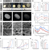
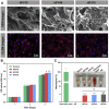
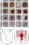
Hemorrhage, infection, and frequent replacement of dressings bring great clinical challenges to wound healing. In this work, Flammulina velutipes extract (FV) and hydroxyethyl cellulose (HEC) were chemically cross-linked and freeze-dried to obtain novel HFV cryogels (named HFVn, with n = 10, 40, or 70 corresponding to the weight percentage of the FV content), which were constructed for wound hemostasis and full-thickness skin defect repair. Systematic characterization experiments were performed to assess the morphology, mechanical properties, hydrophilic properties, and degradation rate of the cryogels. The results indicated that HFV70 showed a loose interconnected-porous structure and exhibited the highest porosity (95%) and water uptake ratio (over 2,500%) with a desirable degradation rate and shape memory properties. In vitro cell culture and hemocompatibility experiments indicated that HFV70 showed improved cytocompatibility and hemocompatibility. It can effectively mimic the extracellular matrix microenvironment and support the adhesion and proliferation of L929 cells, and its hemolysis rate in vitro was less than 5%. Moreover, HFV70 effectively induced tube formation in HUVEC cells in vitro. The results of the bacteriostatic annulus confirmed that HFV70 significantly inhibited the growth of Gram-negative E. coli and Gram-positive S. aureus. In addition, HFV70 showed ideal antioxidant properties, with the DPPH scavenging rate in vitro reaching 74.55%. In vivo rat liver hemostasis experiments confirmed that HFV70 showed rapid and effective hemostasis, with effects comparable to those of commercial gelatin sponges. Furthermore, when applied to the repair of full-thickness skin defects in a rat model, HFV70 significantly promoted tissue regeneration. Histological analysis further confirmed the improved pro-angiogenic and anti-inflammatory activity of HFV70 in vivo. Collectively, our results demonstrated the potential of HFV70 in the treatment of full-thickness skin defects and rapid hemostasis.
Keywords: Flammulina velutipes polysaccharides; cryogel; hemostasis; hydroxyethyl cellulose; wound healing.
Copyright © 2022 Zhu, Chen, Wu, Xiang, Yan, Xie, Tong, Chen and Cai.
Conflict of interest statement
The authors declare that the research was conducted in the absence of any commercial or financial relationships that could be construed as a potential conflict of interest.
Figures


Similar articles
-
Multifunctional chitosan/gelatin@tannic acid cryogels decorated with in situ reduced silver nanoparticles for wound healing.Burns Trauma. 2022 Jul 27;10:tkac019. doi: 10.1093/burnst/tkac019. eCollection 2022. Burns Trauma. 2022. PMID: 35910193 Free PMC article.
-
Multifunctional Tissue-Adhesive Cryogel Wound Dressing for Rapid Nonpressing Surface Hemorrhage and Wound Repair.ACS Appl Mater Interfaces. 2020 Aug 12;12(32):35856-35872. doi: 10.1021/acsami.0c08285. Epub 2020 Jul 29. ACS Appl Mater Interfaces. 2020. PMID: 32805786
-
Green fabrication of seedbed-like Flammulina velutipes polysaccharides-derived scaffolds accelerating full-thickness skin wound healing accompanied by hair follicle regeneration.Int J Biol Macromol. 2021 Jan 15;167:117-129. doi: 10.1016/j.ijbiomac.2020.11.154. Epub 2020 Nov 26. Int J Biol Macromol. 2021. PMID: 33249152
-
Natural Flammulina velutipes-Based Nerve Guidance Conduit as a Potential Biomaterial for Peripheral Nerve Regeneration: In Vitro and In Vivo Studies.ACS Biomater Sci Eng. 2021 Aug 9;7(8):3821-3834. doi: 10.1021/acsbiomaterials.1c00304. Epub 2021 Jul 23. ACS Biomater Sci Eng. 2021. PMID: 34297535
-
Advances in the extraction, purification, structural-property relationships and bioactive molecular mechanism of Flammulina velutipes polysaccharides: A review.Int J Biol Macromol. 2021 Jan 15;167:528-538. doi: 10.1016/j.ijbiomac.2020.11.208. Epub 2020 Dec 2. Int J Biol Macromol. 2021. PMID: 33278442 Review.
References
-
- Anjugam M., Iswarya A., Vaseeharan B. (2016). Multifunctional role of β-1, 3 glucan binding protein purified from the haemocytes of blue swimmer crab Portunus pelagicus and in vitro antibacterial activity of its reaction product. Fish shellfish Immunol. 48, 196–205. 10.1016/j.fsi.2015.11.023 - DOI - PubMed
-
- Begum M. (2014). Official methods of analysis. 2nd ed. Washington D.C.: Association of Official Agricultural Chemists, 832. AOAC1975.
LinkOut - more resources
Full Text Sources

