. 2022 Oct 11:13:967567.
doi: 10.3389/fmicb.2022.967567. eCollection 2022.

CodY is modulated by YycF and affects biofilm formation in Staphylococcus aureus

Affiliations

CodY is modulated by YycF and affects biofilm formation in Staphylococcus aureus

Shizhou Wu et al. Front Microbiol. .

Abstract

Background: Staphylococcus aureus (S. aureus) is the leading cause of various infective diseases, including topical soft tissue infections. The goals of this study were to investigate the roles of YycF and CodY in the regulation of biofilm formation and pathogenicity.

Methods: Electrophoretic mobility shift assay (EMSA) was conducted to validate the bound promoter regions of YycF protein. We constructed the codY up-regulated or down-regulated S. aureus mutants. The biofilm biomass was determined by crystal violet microtiter assay and scanning electron microscopy (SEM). Quantitative RT-PCR analysis was used to detect the transcripts of biofilm-related genes. The live and dead cells of S. aureus biofilm were also investigated by confocal laser scanning microscopy (CLSM). We constructed an abscess infection in Sprague Dawley (SD) rat models to determine the effect of CodY on bacterial pathogenicity. We further used the RAW264.7, which were cocultured with S. aureus, to evaluate the effect of CodY on macrophages apoptosis.

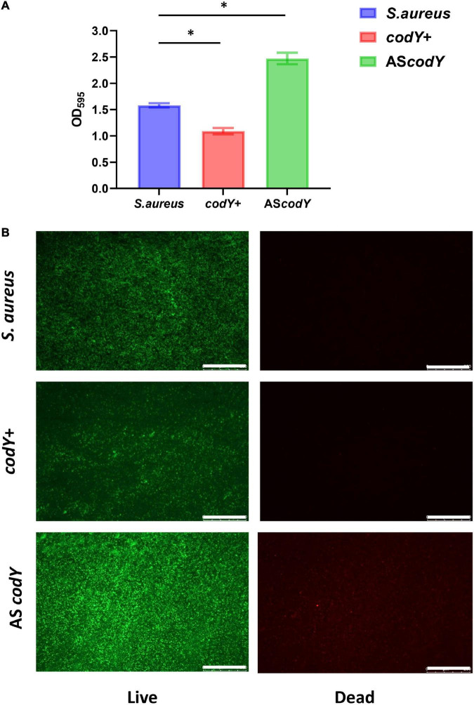
Result: Quantitative RT-PCR analyses reveled that YycF negatively regulates codY expression. EMSA assays indicated that YycF protein directly binds to the promoter regions of codY gene. Quantitative RT-PCR confirmed the construction of dual- mutant stains codY + ASyycF and codY-ASyycF. The SEM results showed that the biofilm formation in the codY + ASyycF group was sparser than those in the other groups. The crystal violet assays indicated that the codY + ASyycF group formed less biofilms, which was consistent with the immunofluorescence results of the lowest live cell ration in the codY + ASyycF group. The expression levels of biofilm-associated icaA gene were significantly reduced in the codY + strain, indicating codY negatively regulates the biofilm formation. Furthermore, CodY impedes the pathogenicity in a rat-infection model. After cocultured with bacteria or 4-h in vitro, the apoptosis rates of macrophage cells were lowest in the codY + group.

Conclusions: YycF negatively regulate the expression of codY. By interaction with codY, YycF could modulate S. aureus biofilm formation via both eDNA- dependent and PIA- dependent pathways, which can be a significant target for antibiofilm. CodY not only impedes the pathogenicity but also has a role on immunoregulation. Thus, the current evidence may provide a supplementary strategy for managing biofilm infections.

Keywords: CodY; Staphylococcus aureus; YycFG; antisense; biofilm formation.


Conflict of interest statement

The authors declare that the research was conducted in the absence of any commercial or financial relationships that could be construed as a potential conflict of interest.

Figures

FIGURE 1
FIGURE 1
YycF negatively regulates codY expression. (A) Consensus YycF binding motif and candidate sequences in promoters of codY. TGTWAH-NNNNN-TGTWAH, where W is A/T and H is A/T/C; EMSA in which promoter regions were obtained by PCR and FAM-labeled. As the negative control, a DNA fragment the same size as the promoter region and similar AT: GC mole ratio, but missing the YycF consensus binding sequence, was used to rule out non-specific binding. (B) Quantitative RT-PCR analysis showed the gene transcripts in S. aureus, and ASyycF strains. S. aureus gene expression was relatively quantified by RT-PCR using 16S as an internal control (n = 5, *P < 0.05).
FIGURE 2
FIGURE 2
CodY interaction with yycF affects biofilm morphology. (A) Quantitative RT-PCR analysis showed the gene expressions in S. aureus, codY + ASyycF, and codY-ASyycF dual- mutant stains. S. aureus gene expression was relatively quantified by RT-PCR using 16S as an internal control (n = 5, *P < 0.05). (B) SEM images of S. aureus, codY + ASyycF, and codY-ASyycF dual- mutant stains.
FIGURE 3
FIGURE 3
CodY interaction with yycF regulates the biofilm biomass. (A) Biomass of S. aureus, codY + ASyycF, and codY-ASyycF dual- mutant stains were quantified by crystal violet staining. Optical densities at 595 nm were measured (n = 5, *P < 0.05). (B) The CLSM observations of S. aureus, codY + ASyycF and codY-ASyycF dual- mutant stains. Green, viable bacteria (SYTO 9); red, dead bacteria (PI); scale bars, 250 μm.
FIGURE 4
FIGURE 4
CodY suppressed biofilm morphology and biofilm-associated genes. (A) Quantitative RT-PCR analysis showed the gene expressions in S. aureus, codY + and codY- mutant stains. S. aureus gene expression was relatively quantified by RT-PCR using 16S as an internal control (n = 5, *P < 0.05). (B) SEM images of S. aureus, codY + and codY- mutant stains.
FIGURE 5
FIGURE 5
CodY decreases the biomass of S. aureus biofilm. (A) Biomass of S. aureus, codY + and codY- mutant stains were quantified by crystal violet staining. Optical densities at 595 nm were measured (n = 5, *P < 0.05). (B) The CLSM observations of S. aureus, codY + and codY- mutant stains. Green, viable bacteria (SYTO 9); red, dead bacteria (PI); scale bars, 250 μm.
FIGURE 6
FIGURE 6
CodY impedes pathogenicity in a rat-infection model. (A) Gross observation of the abscess in S. aureus, codY + and codY- infected group; scale bars, 1 cm. (B) HE staining of the abscess lesions in S. aureus, codY + and codY- infected group; scale bars, 200 μm. (C) The apoptosis rates of macrophage cells when co-cultured with S. aureus, codY + and codY- respectively were measured by the flow cytometer.
FIGURE 7
FIGURE 7
Working model of CodY interactions with YycF affects biofilm formation in Staphylococcus aureus.

References

    1. Augagneur Y., King A. N., Germain-Amiot N., Sassi M., Fitzgerald J. W., Sahukhal G. S., et al. (2020). Analysis of the CodY RNome reveals RsaD as a stress-responsive riboregulator of overflow metabolism in Staphylococcus aureus. Mol. Microbiol. 113 309–325. 10.1111/mmi.14418 - DOI - PubMed
    1. Barbieri G., Voigt B., Albrecht D., Hecker M., Albertini A. M., Sonenshein A. L., et al. (2015). CodY regulates expression of the Bacillus subtilis extracellular proteases Vpr and Mpr. J. Bacteriol. 197 1423–1432. 10.1128/JB.02588-14 - DOI - PMC - PubMed
    1. Bleul L., Francois P., Wolz C. (2021). Two-component systems of S. aureus: Signaling and sensing mechanisms. Genes 13:34. - PMC - PubMed
    1. Brinsmade S. R. (2017). CodY, a master integrator of metabolism and virulence in Gram-positive bacteria. Curr. Genet. 63 417–425. 10.1007/s00294-016-0656-5 - DOI - PubMed
    1. Cardiff R. D., Miller C. H., Munn R. J. (2014). Manual hematoxylin and eosin staining of mouse tissue sections. Cold Spring Harb. Protoc. 2014 655–658. 10.1101/pdb.prot073411 - DOI - PubMed

LinkOut - more resources