

doi: 10.3348/kjr.2022.0617.
Nutcracker Phenomenon and Syndrome May Be More Prevalent Than Previously Thought
Affiliations
- PMID: 36305049
- PMCID: PMC9614295
- DOI: 10.3348/kjr.2022.0617
Item in Clipboard
Nutcracker Phenomenon and Syndrome May Be More Prevalent Than Previously Thought
Korean J Radiol.
2022 Nov.
No abstract available
Conflict of interest statement
The authors have no potential conflicts of interest to disclose.
Figures
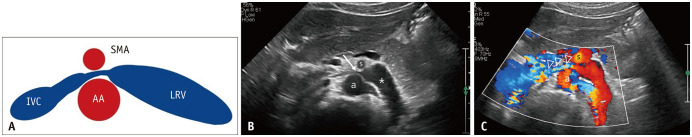
A. Schematic diagram showing the compression of the LRV in the narrow space between the AA and SMA. B, C. Type 1 compression of the LRV in a 33-year-old female with proteinuria. Greyscale (B) and color Doppler US (C) images show a tightly compressed LRV (arrow) between the AA (a) and SMA (s) and a dilated proximal LRV (asterisk). Note the bright-colored jetting of blood flow (arrowheads) from the aortomesenteric LRV. Supplementary video 1 is a video-clip of color Doppler US (C). AA = abdominal aorta, IVC = inferior vena cava, LRV = left renal vein, NCP = nutcracker phenomenon, SMA = superior mesenteric artery, US = ultrasound
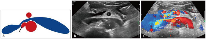
A. Schematic diagram showing the compression of the LRV posteriorly by the origin of the right renal artery (arrow). B, C. Type 2 compression of the LRV in a 68-year-old male with proteinuria. Greyscale (B) and color Doppler US (C) images of the LRV show that the space between the AA (a) and SMA (s) is not narrow, but the LRV is compressed posteriorly by the origin of the right renal artery (arrows). Note the bright-colored jetting of the blood flow (arrowheads) from the site where the right renal artery compresses the LRV. Supplementary video 2 is a video-clip of color Doppler US (C). AA = abdominal aorta, LRV = left renal vein, SMA = superior mesenteric artery, US = ultrasound
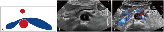
A. Schematic diagram of the LRV narrowed and stretched anterior to the AA. B, C. Type 3 compression of the LRV in a 74-year-old male with microscopic hematuria and proteinuria. Greyscale (B) and color Doppler US (C) images of the LRV show that the LRV is narrowed and stretched over the AA (a). Note the bright-colored jetting of the blood flow (arrowheads) from the site where the LRV is narrowed by stretching over the AA. In addition, scanty flow signals in the dilated proximal LRV indicate slow flow (asterisk). Supplementary video 3 is a video-clip of color Doppler US (C). AA = abdominal aorta, LRV = left renal vein, US = ultrasound
References
-
- Orphanet. Renal nutcracker syndrome. orpha.net Web site. [Accessed September 30, 2022]. https://www.orpha.net/consor/cgi-bin/OC_Exp.php?Lng=GB&Expert=71273 .
-
- Kim SH, Yoon T, Kang E. Nutcracker phenomenon and nutcracker syndrome, one-year experience at a nephrology-uroradiology clinic. ksum.or.kr Web site. [Accessed September 30, 2022]. https://2020.ksum.or.kr/file/KSUM_2020_Program_Abstract_Book.pdf .
Publication types
MeSH terms
LinkOut - more resources
Full Text Sources
