. 2022 Oct 27;185(22):4067-4081.e21.
doi: 10.1016/j.cell.2022.09.026.

Structural basis for Cas9 off-target activity

Affiliations

Structural basis for Cas9 off-target activity

Martin Pacesa et al. Cell. .

Abstract

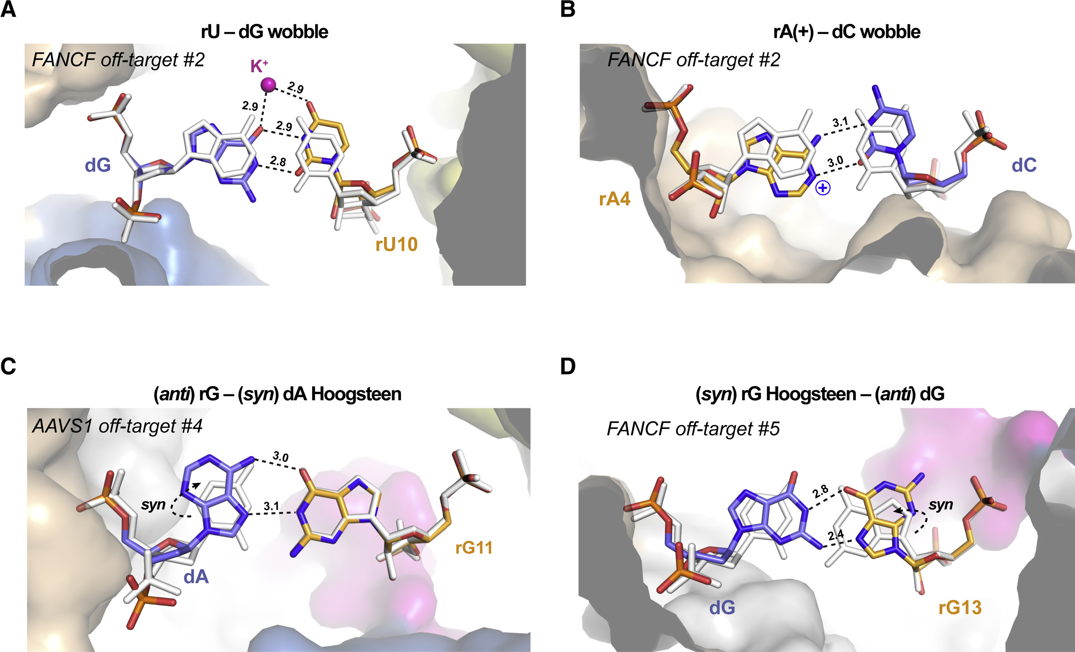
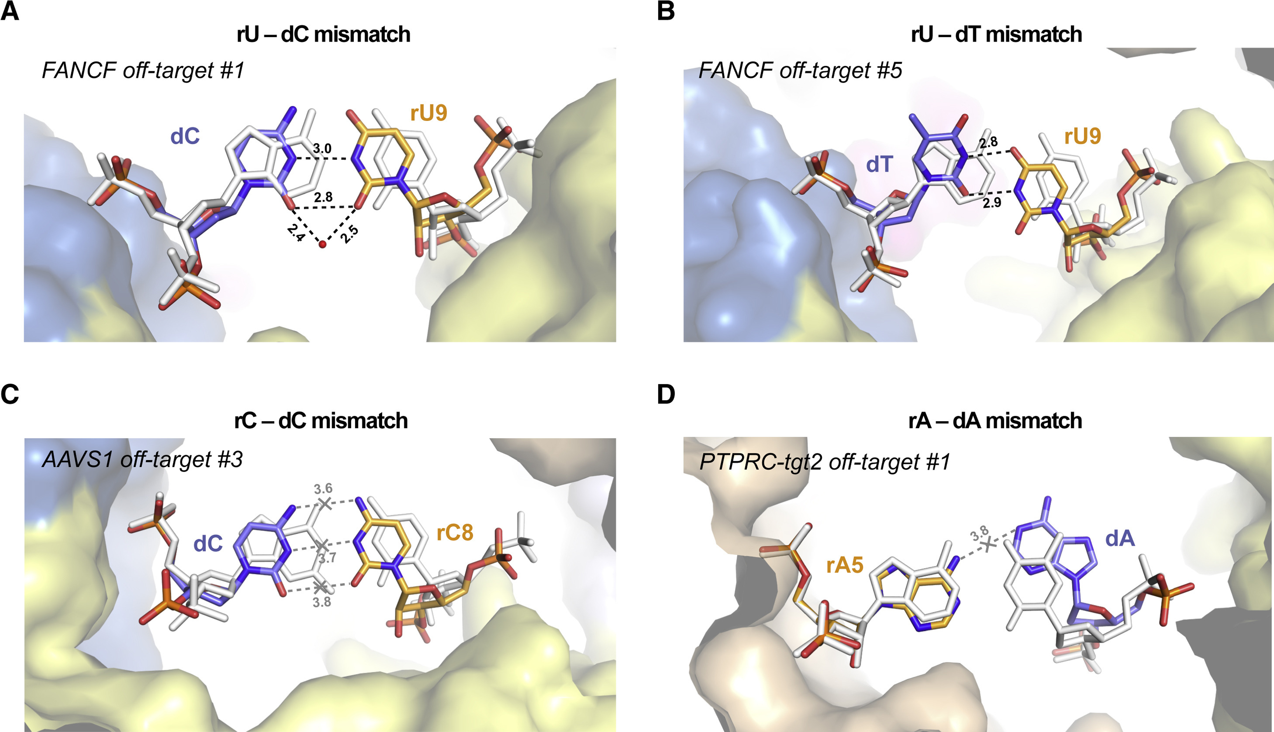
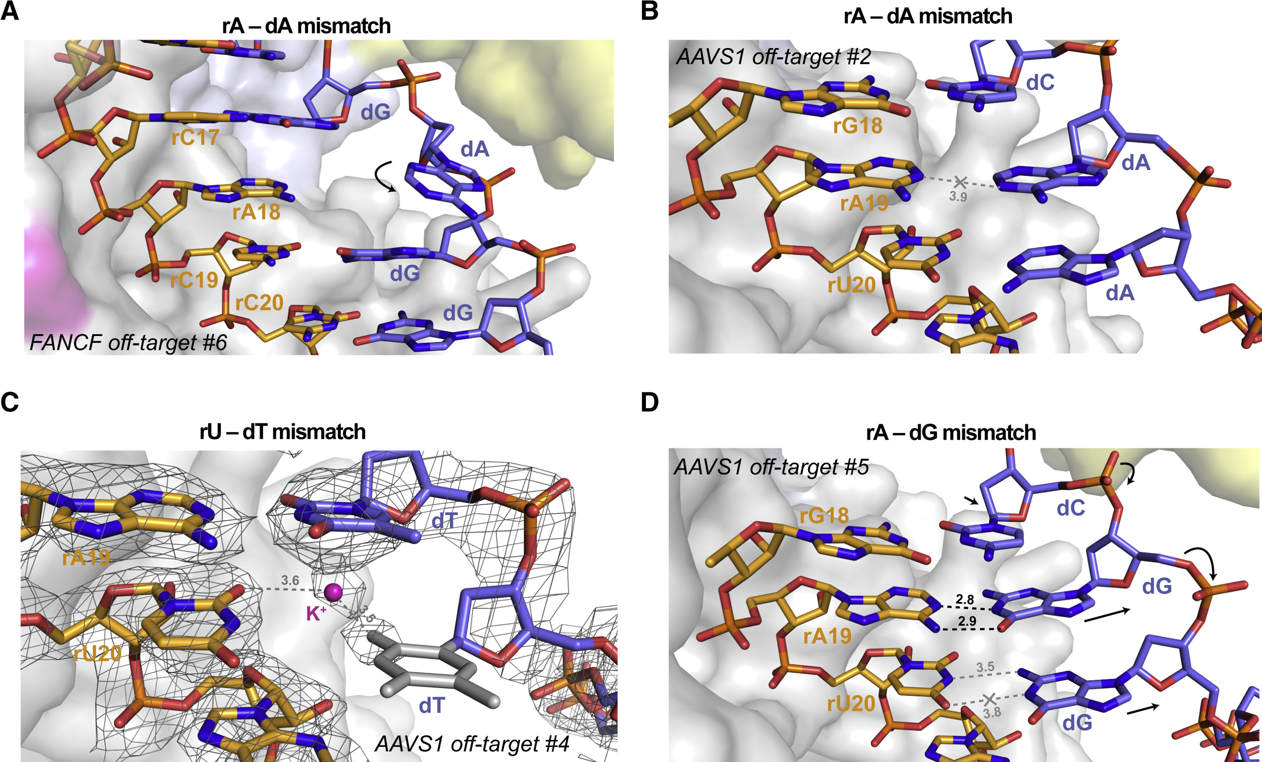
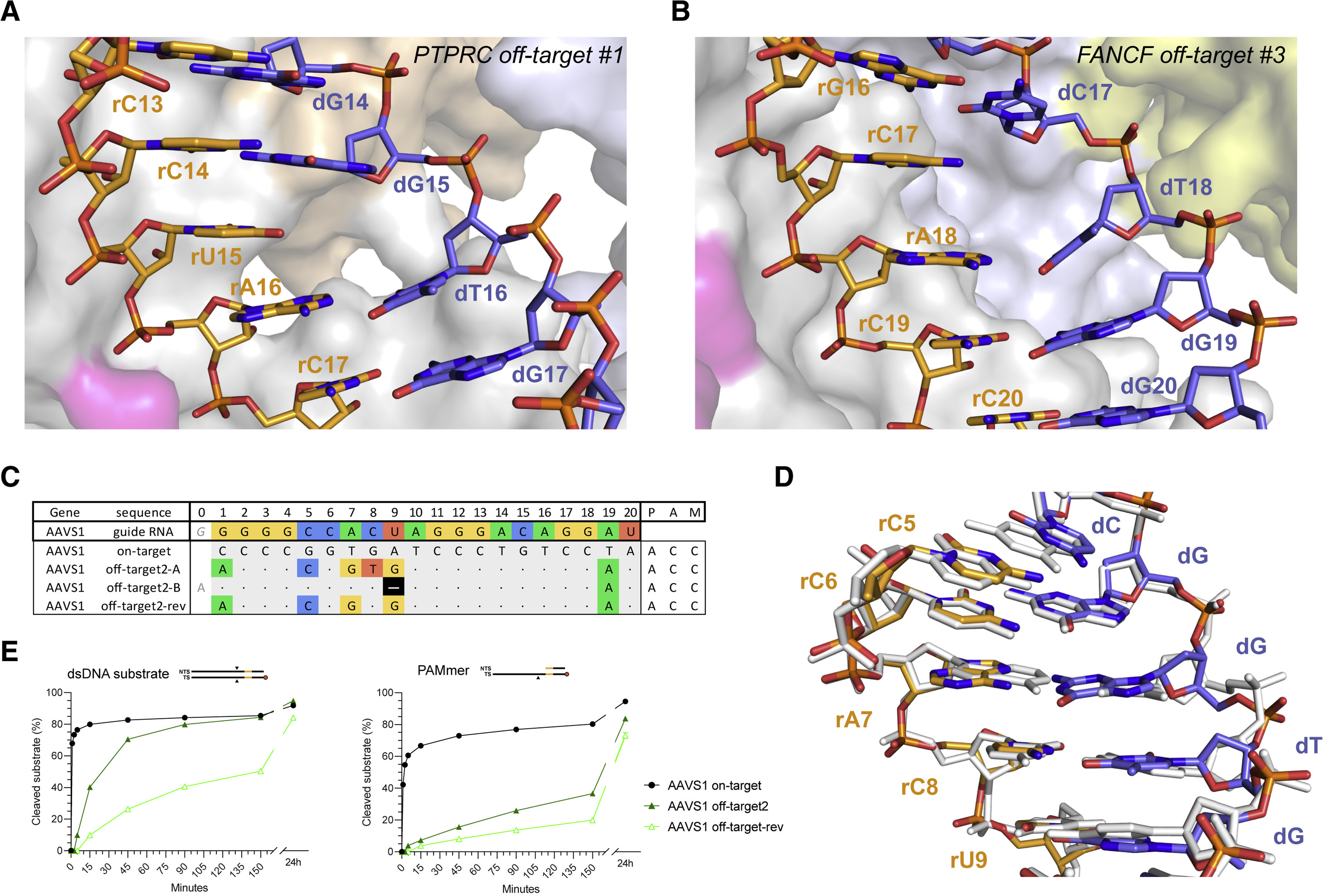
The target DNA specificity of the CRISPR-associated genome editor nuclease Cas9 is determined by complementarity to a 20-nucleotide segment in its guide RNA. However, Cas9 can bind and cleave partially complementary off-target sequences, which raises safety concerns for its use in clinical applications. Here, we report crystallographic structures of Cas9 bound to bona fide off-target substrates, revealing that off-target binding is enabled by a range of noncanonical base-pairing interactions within the guide:off-target heteroduplex. Off-target substrates containing single-nucleotide deletions relative to the guide RNA are accommodated by base skipping or multiple noncanonical base pairs rather than RNA bulge formation. Finally, PAM-distal mismatches result in duplex unpairing and induce a conformational change in the Cas9 REC lobe that perturbs its conformational activation. Together, these insights provide a structural rationale for the off-target activity of Cas9 and contribute to the improved rational design of guide RNAs and off-target prediction algorithms.

Keywords: CRISPR; Cas9; X-ray crystallography; base pairing; genome editing; guide RNA; mismatch; nuclease; off-target.


Conflict of interest statement

Declaration of interests P.D.D. is a current employee of Caribou Biosciences, Inc., and C.-H.L., M.J.I., and P.C. are former employees of Caribou Biosciences, Inc. M.J. is a cofounder of Caribou Biosciences, Inc. M.J., M.J.I., P.C., and P.D.D. are named inventors on patents and patent applications related to CRISPR-Cas technologies. SITE-Seq is a registered trademark of Caribou Biosciences, Inc.

Figures

Figure 1.
Figure 1.. Biochemical and structural analysis of Cas9 off-targets.
(A) Guide RNA and (off-)target DNA sequences selected for biochemical and structural analysis. Matching bases in off-targets are denoted by a dot; nucleotide mismatches and deletions (–) are highlighted. (B) Top: Schematic representation of the guide RNA (orange), TS (blue), and NTS (black) sequences used for crystallisation. The PAM sequence in the DNA is highlighted in yellow. Bottom: Structure of the Cas9 FANCF on-target complex. Individual Cas9 domains are coloured according to the legend; substrate DNA target strand (TS) is coloured blue, non-target strand (NTS) black, and the guide RNA orange.
Figure 2.
Figure 2.. Cas9 off-target binding is enabled by non-canonical base pairing.
Close-up views of (A) rU-dG wobble base pair at duplex position 10 in FANCF off-target #2 complex, (B) rA-dC wobble base pair at position 4 in FANCF off-target #2 complex, (C) rG-dA Hoogsteen base pair at duplex position 11 in AAVS1 off-target #4 complex and (D) rG-dG Hoogsteen base pair at duplex position 13 in FANCF off-target #5 complex. Hydrogen bonding interactions are indicated with dashed lines. Numbers indicate interatomic distances in Å. Corresponding on-target Watson-Crick base pairs are shown in white. Dashed arrows indicate anti-syn isomerization of the dA and rG bases to enable Hoogsteen-edge base pairing. A bound monovalent ion, modelled as K+, is depicted as a purple sphere. See also Figures S7, S8, S9.
Figure 3.
Figure 3.. Duplex backbone distortions facilitate formation of non-canonical base pairs.
(A) Close-up view of the rU-dC base pair at duplex position 9 in FANCF off-target #1 complex, facilitated by lateral displacement of the guide RNA backbone. (B) Zoomed-in view of the rU-dT base pair at position 9 in FANCF off-target #5 complex. (C) Zoomed-in view of the rC-dC mismatch at duplex position 8 in AAVS1 off-target #3 complex. The distances between the cytosine bases indicate lack of hydrogen bonding. (D) Zoomed-in view of the rA-dA mismatch at duplex position 5 in PTPRC-tgt2 off-target #1 complex. Hydrogen bonding interactions are indicated with dashed lines. Corresponding on-target Watson-Crick base pairs are shown in white (for PTPRC-tgt2, the FANCF on-target structure was used and bases were mutated in silico). Numbers indicate interatomic distances in Å. Bound water molecule is depicted as red sphere. See also Figures S10, S11, S12, S13.
Figure 4.
Figure 4.. TS distortion facilitates mismatch accommodation in the seed region of the guide–off-target heteroduplex.
(A) Close-up view of the rA-dA mismatch at position 18 in FANCF off-target #6 complex, showing major groove extrusion of the dA base. (B) Close-up view of the rA-dA mismatch at position 19 in AAVS1 off-target #2 complex, showing retention of the dA base in the duplex stack. (C) Zoomed-in view of the rA-dG base pair at position 19 and the unpaired rU-dG mismatch at position 20 in the AAVS1 off-target #5 complex. (D) Close-up rU-dT mismatch at the PAM-proximal position 20 in AAVS1 off-target #4 complex. Residual electron density indicates the presence of an ion or solvent molecule. Refined 2mFo−DFc electron density map of the heteroduplex, contoured at 1.5σ, is rendered as a grey mesh. Structurally disordered thymine nucleobase for which no unambiguous density is present is in grey. Arrows indicate conformational changes in the TS backbone relative to the on-target complex. See also Figure S14.
Figure 5.
Figure 5.. Off-targets with single-nucleotide deletions are accommodated by base skipping or multiple consecutive mismatches.
(A) Zoomed-in view of the base skip at duplex position 15 in the PTPRC-tgt2 off-target #1 complex. (B) Zoomed-in view of the base skip at duplex position 17 in the FANCF off-target #3 complex. (C) Schematic depiction of alternative base pairing interactions in the AAVS1 off-target #2 complex. AAVS1 off-target #2-rev substrate was designed based on the AAVS1 off-target #2, with the reversal of a single mismatch in the consecutive region back to the corresponding canonical base pair. (D) Structural overlay of the AAVS1 off-target #2 (coloured) and AAVS1 on-target (white) heteroduplexes. (E) Cleavage DNA kinetics of AAVS1 on-target, off-target #2 and off-target #2-rev substrates. See also Figures S15, S16, S17, S18, S19.
Figure 6.
Figure 6.. FANCF off-target #4 exhibits conformational changes in the REC2/3 and HNH domains due to PAM-distal duplex unpairing.
(A) Close-up view of the unpairing of mismatched bases at the PAM-distal end of the FANCF off-target #4 heteroduplex. The last two nucleotides on each strand could not be modelled due to structural disorder. (B) Overlay of the FANCF off-target #4 and FANCF on-target complex structures. The FANCF off-target #4 complex is coloured according to the domain legend in Figure 1A, FANCF on-target complex is shown in white. The REC1, RuvC, and PAM-interaction domains have been omitted for clarity, as no structural differences are observed. See also Figures S20, S21.

Similar articles

Cited by

References

    1. Abe N, Dror I, Yang L, Slattery M, Zhou T, Bussemaker HJ, Rohs R, and Mann RS (2015). Deconvolving the recognition of DNA shape from sequence. Cell 161, 307–318. - PMC - PubMed
    1. Adams PD, Afonine PV, Bunkoczi G, Chen VB, Davis IW, Echols N, Headd JJ, Hung LW, Kapral GJ, Grosse-Kunstleve RW, et al. (2010). PHENIX: a comprehensive Python-based system for macromolecular structure solution. Acta Crystallogr D Biol Crystallogr 66, 213–221. - PMC - PubMed
    1. Afek A, Shi H, Rangadurai A, Sahay H, Senitzki A, Xhani S, Fang M, Salinas R, Mielko Z, Pufall MA, et al. (2020). DNA mismatches reveal conformational penalties in protein-DNA recognition. Nature. - PMC - PubMed
    1. Anders C, Niewoehner O, Duerst A, and Jinek M. (2014). Structural basis of PAM-dependent target DNA recognition by the Cas9 endonuclease. Nature 513, 569–573. - PMC - PubMed
    1. Anzalone AV, Koblan LW, and Liu DR (2020). Genome editing with CRISPR-Cas nucleases, base editors, transposases and prime editors. Nat Biotechnol 38, 824–844. - PubMed

Publication types