Review
. 2023 Oct;193(10):1427-1439.
doi: 10.1016/j.ajpath.2022.08.010. Epub 2022 Oct 25.

Liver Iron Loading in Alcohol-Associated Liver Disease

Affiliations
Review

Liver Iron Loading in Alcohol-Associated Liver Disease

Najma Ali et al. Am J Pathol. 2023 Oct.

Abstract

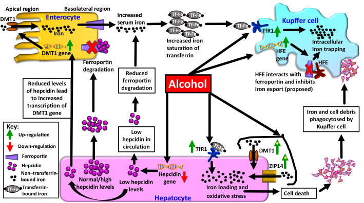
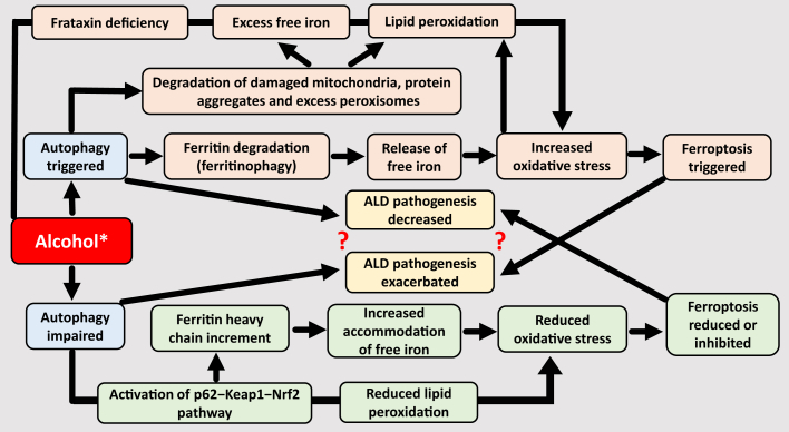
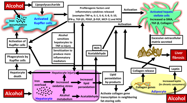
Alcohol-associated liver disease (ALD) is a common chronic liver disease with increasing incidence worldwide. Alcoholic liver steatosis/steatohepatitis can progress to liver fibrosis/cirrhosis, which can cause predisposition to hepatocellular carcinoma. ALD diagnosis and management are confounded by several challenges. Iron loading is a feature of ALD which can exacerbate alcohol-induced liver injury and promote ALD pathologic progression. Knowledge of the mechanisms that mediate liver iron loading can help identify cellular/molecular targets and thereby aid in designing adjunct diagnostic, prognostic, and therapeutic approaches for ALD. Herein, the cellular mechanisms underlying alcohol-induced liver iron loading are reviewed and how excess iron in patients with ALD can promote liver fibrosis and aggravate disease pathology is discussed. Alcohol-induced increase in hepatic transferrin receptor-1 expression and up-regulation of high iron protein in Kupffer cells (proposed) facilitate iron deposition and retention in the liver. Iron is loaded in both parenchymal and nonparenchymal liver cells. Iron-loaded liver can promote ferroptosis and thereby contribute to ALD pathology. Iron and alcohol can independently elevate oxidative stress. Therefore, a combination of excess iron and alcohol amplifies oxidative stress and accelerates liver injury. Excess iron-stimulated hepatocytes directly or indirectly (through Kupffer cell activation) activate the hepatic stellate cells via secretion of proinflammatory and profibrotic factors. Persistently activated hepatic stellate cells promote liver fibrosis, and thereby facilitate ALD progression.


Figures

Figure 1
Figure 1
Cellular events underlying alcohol-induced iron loading in different cell types. Alcohol consumption decreases hepcidin levels in the circulation. In turn, this increases intestinal absorption of iron. Elevated serum iron levels cause iron deposition in various cell types, including the hepatocytes and Kupffer cells in the liver, via elevation in transferrin receptor-1 (TfR1) and high iron protein (HFE; proposed). Also, alcohol-induced elevations of non–transferrin-bound iron (NTBI) transporters zinc-regulated, iron-regulated transporter-like protein (ZIP) and divalent metal-ion transporter 1 (DMT1) on hepatocytes, as observed in some studies, aid in hepatocyte iron loading. Green arrows with yellow stars indicate variability in results with regard to alcohol-induced elevation of these NTBI transporters.
Figure 2
Figure 2
Putative associations between autophagy, ferritinophagy, and ferroptosis in the presence of alcohol. Alcohol has a dual effect of autophagy (ie, it can both stimulate and impair autophagy). This differential effect of alcohol on autophagy and the consequent ambiguity is indicated through an asterisk in the figure. Degradation of ferritin via autophagy is ferritinophagy. Ferritinophagy can trigger ferroptosis, whereas increment in ferritin can increase the probability of accommodating free iron, thereby reducing excess iron–induced oxidative stress, and consequently reducing ferroptosis. Autophagy can also trigger ferroptosis through ferritinophagy-independent routes, such as those involving frataxin deficiency, and degradation of damaged or excess cellular components that eventually increases free iron levels and/or lipid peroxidation., Interestingly, although autophagy can trigger ferroptosis, which can exacerbate alcohol-associated liver disease (ALD) pathology, autophagy appears to also impart a protective effect and decrease or blunt ALD pathology. These apparently opposing concepts have been indicated by question marks in the figure and need further clarification.
Figure 3
Figure 3
Intercellular events depicting the role of iron in enhancing alcohol-induced liver fibrosis. Alcohol can cause iron loading in the hepatocytes and Kupffer cells. Oxidative injury to hepatocytes due to excess iron and alcohol can lead to hepatocyte death. Kupffer cells phagocytose dead/damaged hepatocytes and get activated. Activated Kupffer cells release profibrotic cytokines and activate the hepatic stellate cells (HSCs). In addition, profibrotic/inflammatory cytokines released from injured hepatocytes together with reactive oxygen species (ROS) and acetaldehyde produced from alcohol metabolism in the hepatocytes activate the HSCs. Following activation, HSCs secrete profibrotic factors and excessive extracellular matrix that collectively form the basis for liver fibrosis. Adipocytes also play a role in promoting alcohol-induced liver fibrosis, and together with excess iron, the pathology may be aggravated. β-FGF, β-fibroblast growth factor; IFN-γ, interferon-γ; MCP-1, monocyte chemoattractant protein-1; PDGF, platelet-derived growth factor; α-SMA, α-smooth muscle actin; TGF, transforming growth factor; TNF-α, tumor necrosis factor-α.

Comment in

Similar articles

Cited by

References

    1. Manthey J., Shield K.D., Rylett M., Hasan O.S.M., Probst C., Rehm J. Global alcohol exposure between 1990 and 2017 and forecasts until 2030: a modelling study. Lancet. 2019;393:2493–2502. - PubMed
    1. Rehman A., Mehta K.J. Betaine in ameliorating alcohol-induced hepatic steatosis. Eur J Nutr. 2022;61:1167–1176. - PMC - PubMed
    1. Harrison-Findik D.D. Role of alcohol in the regulation of iron metabolism. World J Gastroenterol. 2007;13:4925–4930. - PMC - PubMed
    1. Ganne-Carrié N., Christidis C., Chastang C., Ziol M., Chapel F., Imbert-Bismut F., Trinchet J.C., Guettier C., Beaugrand M. Liver iron is predictive of death in alcoholic cirrhosis: a multivariate study of 229 consecutive patients with alcoholic and/or hepatitis C virus cirrhosis: a prospective follow up study. Gut. 2000;46:277–282. - PMC - PubMed
    1. Eng S.C., Taylor S.L., Reyes V., Raaka S., Berger J., Kowdley K.V. Hepatic iron overload in alcoholic end-stage liver disease is associated with iron deposition in other organs in the absence of HFE-1 hemochromatosis. Liver Int. 2005;25:513–517. - PubMed