

Discovery and functional characterization of the oncogenicity and targetability of a novel NOTCH1-ROS1 gene fusion in pediatric angiosarcoma
- PMID: 36307212
- PMCID: PMC9632357
- DOI: 10.1101/mcs.a006222
Discovery and functional characterization of the oncogenicity and targetability of a novel NOTCH1-ROS1 gene fusion in pediatric angiosarcoma
Abstract
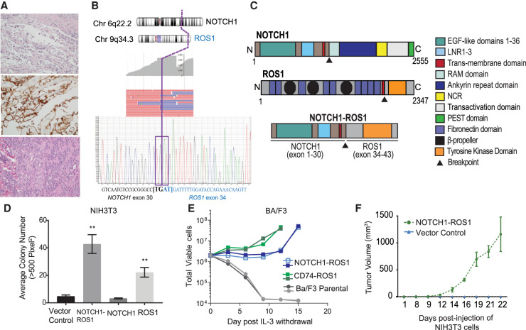
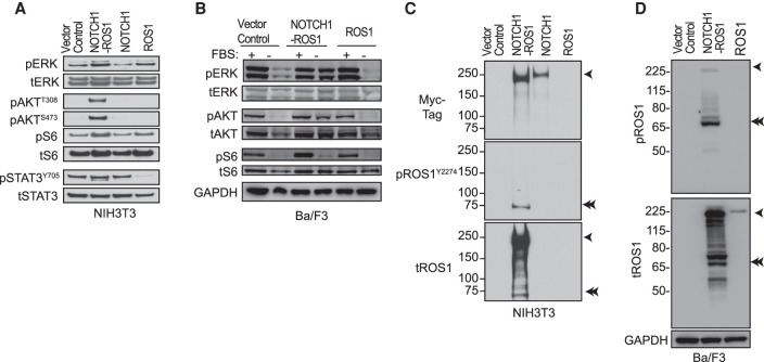
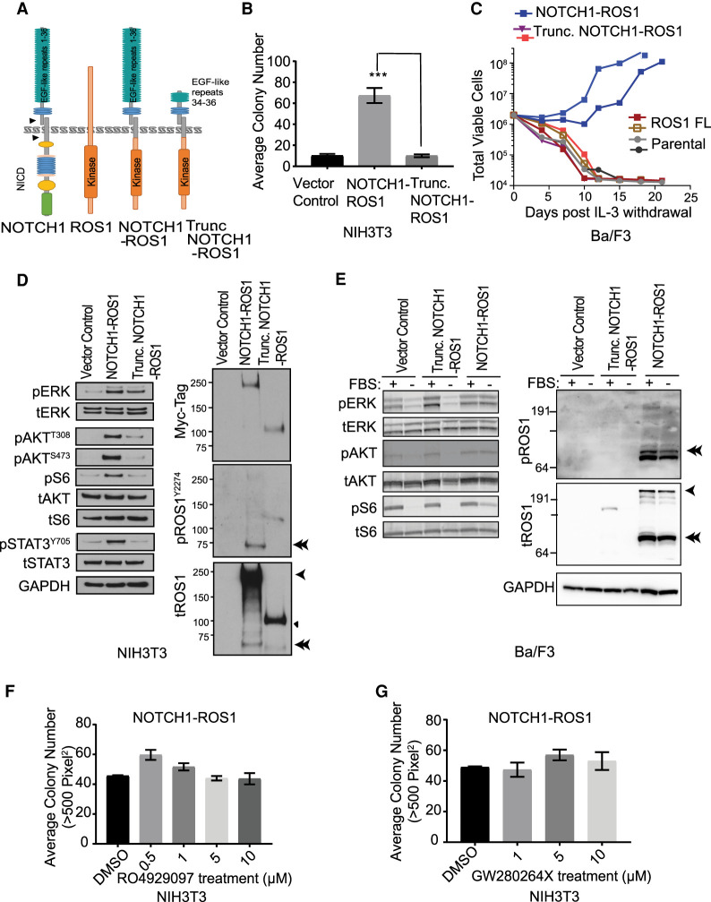
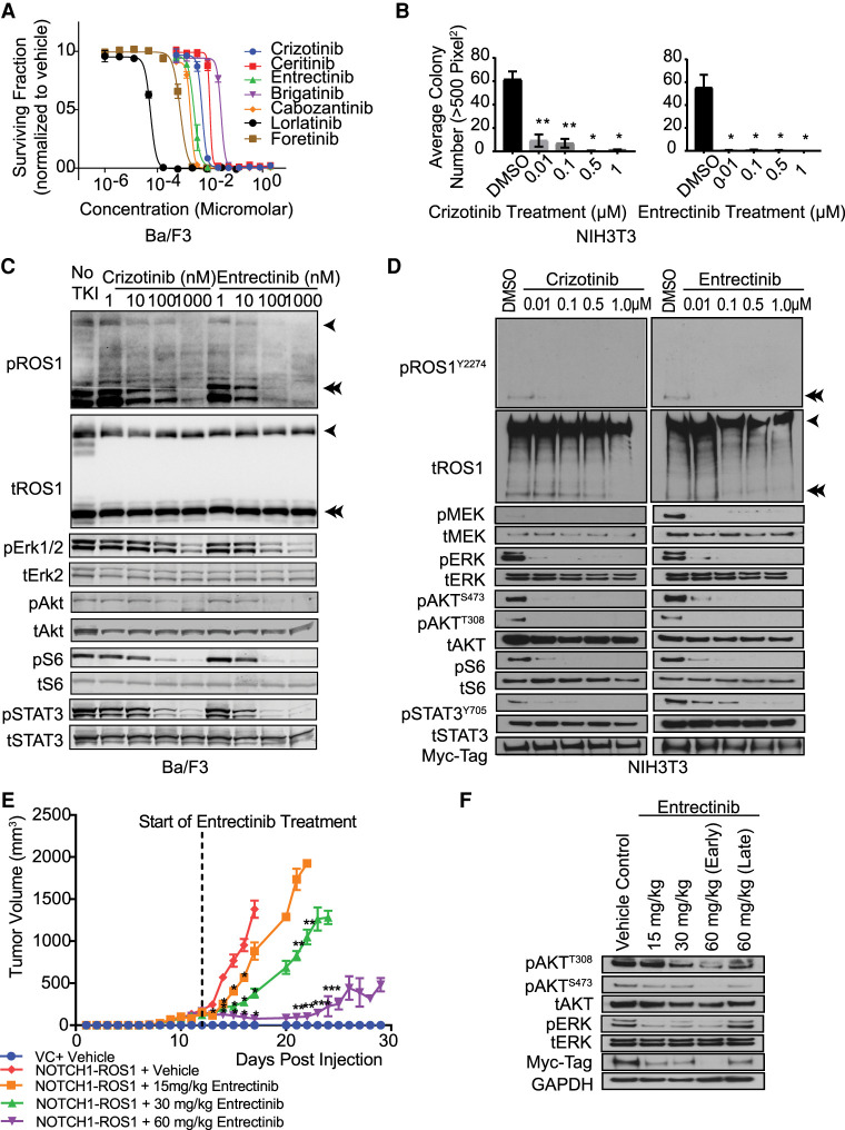
Angiosarcomas are rare, malignant soft tissue tumors in children that arise in a wide range of anatomical locations and have limited targeted therapies available. Here, we report a rare case of a pediatric angiosarcoma (pAS) with Li-Fraumeni syndrome (LFS) expressing a novel NOTCH1-ROS1 gene fusion. Although both NOTCH1 and ROS1 are established proto-oncogenes, our study is the first to describe the mechanistic role of NOTCH1-ROS1 fusion arising via intrachromosomal rearrangement. NOTCH1-ROS1 displayed potent neoplastic transformation propensity in vitro, and harbors tumorigenic potential in vivo, where it induced oncogenic activation of the MAPK, PI3K/mTOR, and JAK-STAT signaling pathways in a murine allograft model. We found an unexpected contribution of the NOTCH1 extracellular region in mediating NOTCH1-ROS1 activation and oncogenic function, highlighting the contribution of both NOTCH1 and ROS1 fusion partners in driving tumorigenicity. Interestingly, neither membrane localization nor fusion protein dimerization were found to be essential for NOTCH1-ROS1 fusion oncogenicity. To target NOTCH1-ROS1-driven tumors, we tested both NOTCH1-directed inhibitors and ROS1-targeted tyrosine kinase inhibitors (TKI) in heterologous models (NIH3T3, Ba/F3). Although NOTCH1 inhibitors did not suppress NOTCH1-ROS1-driven oncogenic growth, we found that oral entrectinib treatment effectively suppressed the growth of NOTCH-ROS1-driven tumors. Taken together, we report the first known pAS case with a novel NOTCH1-ROS1 alteration along with a detailed report on the function and therapeutic targeting of NOTCH1-ROS1. Our study highlights the importance of genomic profiling of rare cancers such as pAS to reveal actionable drivers and improve patient outcomes.
Keywords: metastatic angiosarcoma.
© 2022 Jain et al.; Published by Cold Spring Harbor Laboratory Press.
Figures
Similar articles
-
Identification of a Novel MAN1A1-ROS1 Fusion Gene Through mRNA-based Screening for Tyrosine Kinase Gene Aberrations in a Patient with Leiomyosarcoma.Clin Orthop Relat Res. 2021 Apr 1;479(4):838-852. doi: 10.1097/CORR.0000000000001548. Clin Orthop Relat Res. 2021. PMID: 33196586 Free PMC article.
-
MAPK Pathway Alterations Correlate with Poor Survival and Drive Resistance to Therapy in Patients with Lung Cancers Driven by ROS1 Fusions.Clin Cancer Res. 2020 Jun 15;26(12):2932-2945. doi: 10.1158/1078-0432.CCR-19-3321. Epub 2020 Mar 2. Clin Cancer Res. 2020. PMID: 32122926 Free PMC article.
-
Novel TPR::ROS1 Fusion Gene Activates MAPK, PI3K and JAK/STAT Signaling in an Infant-type Pediatric Glioma.Cancer Genomics Proteomics. 2022 Nov-Dec;19(6):711-726. doi: 10.21873/cgp.20354. Cancer Genomics Proteomics. 2022. PMID: 36316040 Free PMC article.
-
ROS1 protein-tyrosine kinase inhibitors in the treatment of ROS1 fusion protein-driven non-small cell lung cancers.Pharmacol Res. 2017 Jul;121:202-212. doi: 10.1016/j.phrs.2017.04.022. Epub 2017 Apr 30. Pharmacol Res. 2017. PMID: 28465216 Review.
-
Identification of a novel KLC1-ROS1 fusion in a case of pediatric low-grade localized glioma.Brain Tumor Pathol. 2019 Jan;36(1):14-19. doi: 10.1007/s10014-018-0330-3. Epub 2018 Oct 22. Brain Tumor Pathol. 2019. PMID: 30350109 Review.
Cited by
-
Decoding Oncofusions: Unveiling Mechanisms, Clinical Impact, and Prospects for Personalized Cancer Therapies.Cancers (Basel). 2023 Jul 19;15(14):3678. doi: 10.3390/cancers15143678. Cancers (Basel). 2023. PMID: 37509339 Free PMC article. Review.
-
A Novel TEK::GAB2 Gene Fusion in Pediatric Angiosarcoma of Pelvic soft Tissue: A Case Report and Literature Review.Pediatr Dev Pathol. 2025 Jan-Feb;28(1):58-62. doi: 10.1177/10935266241279073. Epub 2024 Sep 9. Pediatr Dev Pathol. 2025. PMID: 39248342 Free PMC article. Review.
-
Imaging features of pediatric angiosarcomas: clinical, pathologic, and radiological review.Pediatr Radiol. 2024 Oct;54(11):1873-1883. doi: 10.1007/s00247-024-06041-0. Epub 2024 Sep 3. Pediatr Radiol. 2024. PMID: 39223383
References
-
- Calvete O, Martinez P, Garcia-Pavia P, Benitez-Buelga C, Paumard-Hernández B, Fernandez V, Dominguez F, Salas C, Romero-Laorden N, Garcia-Donas J, et al. 2015. A mutation in the POT1 gene is responsible for cardiac angiosarcoma in TP53-negative Li–Fraumeni-like families. Nat Commun 6: 8383. 10.1038/ncomms9383 - DOI - PMC - PubMed
Publication types
MeSH terms
Substances
Grants and funding
LinkOut - more resources
Full Text Sources
Molecular Biology Databases
Research Materials
Miscellaneous