. 2022 Oct 28;12(1):18161.
doi: 10.1038/s41598-022-21687-2.

Performance evaluation of a preclinical SPECT/CT system for multi-animal and multi-isotope quantitative experiments

Affiliations

Performance evaluation of a preclinical SPECT/CT system for multi-animal and multi-isotope quantitative experiments

Elena Prieto et al. Sci Rep. .

Abstract

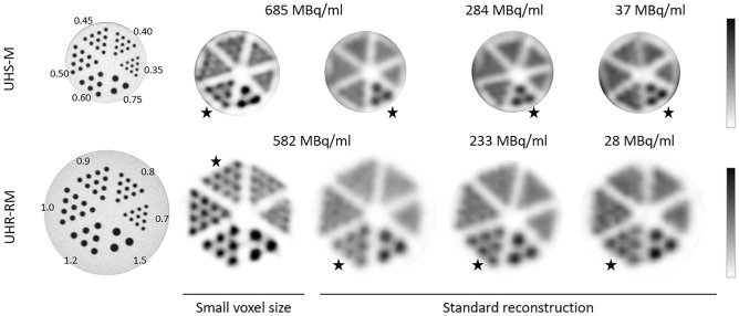
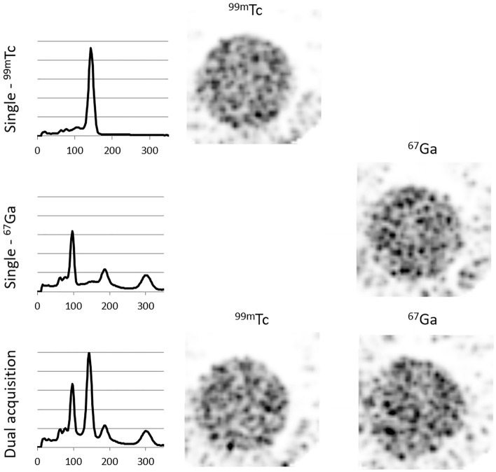
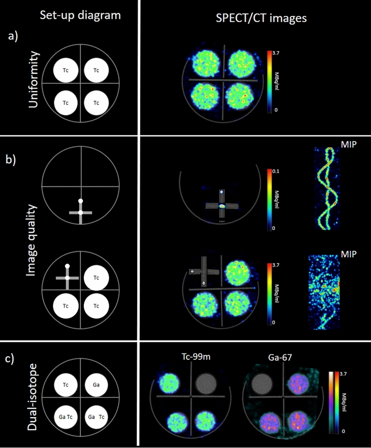
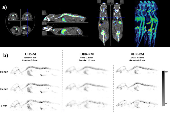
The aim was to study the performance of the U-SPECT6/CT E-class system for preclinical imaging, to later demonstrate the viability of simultaneous multi-animal and multi-isotope imaging with reliable quantitative accuracy. The performance of the SPECT was evaluated for two collimators dedicated for mouse (UHS-M) and rat imaging (UHR-RM) in terms of sensitivity, energy resolution, uniformity and spatial resolution. Point sources, hot‑rod and uniform phantoms were scanned, and additional tests were carried out to evaluate singular settings such as simultaneous multi-isotope acquisition and imaging with a multi-bed system. For in-vivo evaluation, simultaneous triple-isotope and multi-animal studies were performed on mice. Sensitivity for 99mTc was 2370 cps/MBq for the UHS-M collimator and 493 cps/MBq for the UHR-RM. Rods of 0.6 mm and 0.9 mm were discernible with the UHS-M and UHR-RM collimators respectively, with optimized reconstruction. Uniformity in low counting conditions has proven to be poor (> 75%). Multi-isotope and multi-bed phantom acquisitions demonstrated accurate quantification. In mice, simultaneous multi-isotope imaging provided the separate distribution of 3 tracers and image quality of the multi-mouse bone scan was adequate. The U-SPECT6/CT E-class has shown good sensitivity and spatial resolution. This system provides quantitative images with suitable image quality for multi-mouse and multi-isotope acquisitions.


Conflict of interest statement

The authors declare no competing interests.

Figures

Figure 1
Figure 1
Spatial resolution values obtained with the Derenzo phantoms for the UHS-M (up) and UHR-RM (down) collimators. The image on the left shows the CT acquisition of the phantom, whereas SPECT images follow on the right. The first two images represent the same activity concentrations but different reconstruction (minimum voxel size and standard reconstruction). The color bar was adjusted according to the radioactive concentration within the phantom. Stars are used in the figure to mark the minimum distinguishable rod in each case.
Figure 2
Figure 2
Uniformity results obtained for the UHS-M and UHR-RM collimators using different geometries (see Table 1 for detailed description).
Figure 3
Figure 3
Spectra and images obtained with the single (first and second rows) and dual isotope (bottom) acquisitions with the UHS-M collimator.
Figure 4
Figure 4
SPECT/CT images obtained with the multi-bed device and the UHR-RM collimator. The first column shows a schematic representation of the experimental set-up. (a) SPECT axial slice of four uniform syringes, (b) Sinusoidal phantom acquired alone and with three uniform syringes. Fused SPECT/CT axial slices and MIP reconstructions are shown. (c) Syringes with 67Ga and 99mTc for multi-isotope performance evaluation. Fused SPECT/CT images reconstructed for each radionuclide are presented.
Figure 5
Figure 5
Simultaneous SPECT/CT images obtained for three different tracers labeled with 99mTc, 67Ga and 125I. Separate reconstruction has been obtained for each isotope.
Figure 6
Figure 6
Mouse bone scan. (a) Axial, sagittal and coronal slices and MIP reconstruction of SPECT/CT fusion images to show multi-mouse acquisitions from four mice simultaneously. (b) SPECT sagittal slices of a mouse injected with 99mTc-HDP using either the UHS-M or the UHR-RM collimators applying different reconstruction protocols. The 99mTc HDP uptake is clearly detected in the bones (vertebrae, skull) and also in the liver.

References

    1. Lauber DT, et al. State of the art in vivo imaging techniques for laboratory animals. Lab. Anim. 2017;51:465–478. doi: 10.1177/0023677217695852. - DOI - PubMed
    1. Chomet M, et al. Performance of nanoScan PET/CT and PET/MR for quantitative imaging of 18F and 89Zr as compared with ex vivo biodistribution in tumor-bearing mice. EJNMMI Res. 2021;11:1–11. doi: 10.1186/s13550-021-00799-2. - DOI - PMC - PubMed
    1. Franc BL, Acton PD, Mari C, Hasegaway BH. Small-animal SPECT and SPECT/CT: Important tools for preclinical investigation. J. Nucl. Med. 2008;49:1651–1663. doi: 10.2967/jnumed.108.055442. - DOI - PubMed
    1. Jang BS. MicroSPECT and microPET imaging of small animals for drug development. Toxicol. Res. 2013;29:1–6. doi: 10.5487/TR.2013.29.1.001. - DOI - PMC - PubMed
    1. Deleye S, et al. Performance evaluation of small-animal multipinhole μSPECT scanners for mouse imaging. Eur. J. Nucl. Med. Mol. Imaging. 2013;40:744–758. doi: 10.1007/s00259-012-2326-2. - DOI - PubMed

Publication types