. 2022 Oct 28;13(1):6439.
doi: 10.1038/s41467-022-33832-6.

Nebulized fusion inhibitory peptide protects cynomolgus macaques from measles virus infection

Affiliations

Nebulized fusion inhibitory peptide protects cynomolgus macaques from measles virus infection

Olivier Reynard et al. Nat Commun. .

Abstract

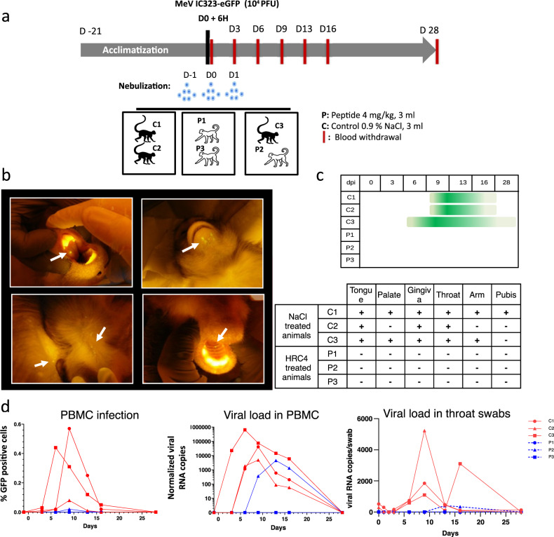
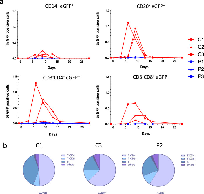
Measles is the most contagious airborne viral infection and the leading cause of child death among vaccine-preventable diseases. We show here that aerosolized lipopeptide fusion inhibitor, derived from heptad-repeat regions of the measles virus (MeV) fusion protein, blocks respiratory MeV infection in a non-human primate model, the cynomolgus macaque. We use a custom-designed mesh nebulizer to ensure efficient aerosol delivery of peptide to the respiratory tract and demonstrate the absence of adverse effects and lung pathology in macaques. The nebulized peptide efficiently prevents MeV infection, resulting in the complete absence of MeV RNA, MeV-infected cells, and MeV-specific humoral responses in treated animals. This strategy provides an additional means to fight against respiratory infection in non-vaccinated people, that can be readily translated to human trials. It presents a proof-of-concept for the aerosol delivery of fusion inhibitory peptides to protect against measles and other airborne viruses, including SARS-CoV-2, in case of high-risk exposure.


Conflict of interest statement

S. Le Guellec is employed by DTF Medical (Saint Etienne, France) and L. Vecellio was employed by DTF Medical from 2001 to 2018 and by Nemera (La Verpilliere, France) from 2018 to 2020. The remaining authors declare no competing interests.

Figures

Fig. 1
Fig. 1. HRC4 lipopeptide treatment does not generate MeV escape variants.
a Schematic presentation of viral passaging. Measles virus MeV IC323-eGFP was serially passaged 8 times on the Vero-hSLAM cells in presence of either 1 µM [FIP-PEG4]2-chol (red) or HRC4 peptide (blue), added to the culture after the infection. Virus was titrated after each passage and 100 Plaque Forming Units (PFU) used for each subsequent infection. Sequencing of viral RNA after the 8th passage revealed two mutations in the F-HRC domain for the infection done in the presence of [FIP-PEG4]2-chol: V459I and N462S, while no mutations in the presence of HRC4 peptide were found. b The most frequent mutation events in F after MeV IC323 was serially passaged 8 times on the Vero-hSLAM cells in presence of [FIP-PEG4]2-chol. c Inhibition of cell-cell fusion mediated by MeV F bearing the indicated mutations by either 5 µM [FIP-PEG4]2-chol or HRC4 peptide, using HEK-293T cells transfected with hSLAM and the omega reporter subunit of β-gal, incubated with cells co-expressing viral glycoproteins (IC323 H and F) and the alpha reporter subunit of β-gal (**** p < 0,0001, Two-Way ANOVA multiple comparison test, n = 3 biologically independent samples in 2 independent experiments). Data are presented as mean values ±SD. Source data are provided as a Source Data file. d Schematic of the HRC4 lipopeptide, used in the further study.
Fig. 2
Fig. 2. Utilization of the prototype mesh nebulizer with low Volume Medium Diameter preserves functional activity of HRC4 lipopeptide.
a Composition of the prototype mesh nebulizer used in experiments: 1. electronic controller; 2. piezo-electric vibrator; 3. reservoir containing the mesh; 4. facemask; 5. one-way inspiratory valve; 6. absolute filter. b Graphical representation of particle size distribution obtained from laser diffraction analysis of aerosolized 3 ml of HRC4 lipopeptide. Plain line presents the mean values of four different nebulizers used in the study and dotted line standard deviation. Percentage of particles <5 µm and <2 µm present the fraction of aerosol below the indicated size, corresponding to the aerosol penetrating into either lung in general (<5 µm) or into alveolar regions of lungs (<2 µm). Volume Mean Diameter (VMD) presents the mean size of generated aerosols. c Fusion inhibitory activity of HRC4, measured before or after peptide nebulization, using β-gal complementation assay. Data represent the mean ± SD of two independent experiments. d Antiviral activity of HRC4 measured prior and after nebulization, determined by IC50 measurement using plaque reduction assay on Vero-hSLAM cells, and cytotoxicity assay, performed by assessing of cell viability 96 h after addition of HRC4 by MTT assay. Data represent the mean ± SD of of three independent experiments (Man-Whitney test, p = 0,8535). Source data are provided as a Source Data file.
Fig. 3
Fig. 3. Delivery of the aerosol into macaques’ lungs.
a Representative scintigraphy imaging of different regions of interest in a cynomolgus monkeys nebulized with 99mTC-DTPA tracer (74 Mbq) in 3 ml NaCl 0.9%, using a prototype mesh nebulizer and measured by E-cam gamma camera. Mean values ±SD of the distribution of aerosol deposition were determined from the digitalized images obtained in four experiments. b Lung localization of the aerosolized HRC4 peptide analyzed in cranial lobe lung sections from monkeys nebulized under mechanical ventilation for the indicated time: 15 min (n = 1), 24 h (n = 2) or 16 h (n = 1), prior to euthanasia. Staining was performed with rabbit anti-HRC peptide and goat anti-rabbit Alexa 555 (orange staining) and DAPI was used to stain nuclei (blue staining) (Scale bar: 20 µm). c Quantification of HRC4 in the NHPs’ serum after peptide nebulization, by ELISA. Histograms present mean values ± SD from 2 independent experiments. Significant increase of HRC4 in the serum, following the nebulization, was confirmed using One way ANOVA (Prism 8.4.3 software, p = 0.0004). d Determination of the presence of anti-HRC4 antibodies (IgG, IgA and IgM) by ELISA in the serum of peptide-nebulized macaques, from either HRC4 biodistribution or MeV-infection study, P1-5 (values obtained at day 0 are subtracted from those at D28 for each individual animal). Negative control (C-) corresponds to serum of a naïve macaque and positive control (C + ) to rabbit anti-HRC4 antiserum. Source data are provided as a Source Data file.
Fig. 4
Fig. 4. Evolution of biochemical and haematological parameters in the blood cynomolgus monkeys following the nebulization of either saline (control) or HRC4 (peptide).
a Concentration of indicated biochemical parameters in blood of monkeys, measured on days 0, 1, 2, 3, 6 and 28 after nebulization of either saline solution (0.9% NaCl, n = 3) or HRC4 peptide (4 mg/kg, n = 5); b Haematological parameters measured at days 0, 1, 2, 3, 6 and 28 after nebulisation of either saline solution (0.9% NaCl, n =3) or HRC4 peptide (4 mg/kg, n = 5). ALAT Alanine aminotransferase, ASAT Aspartate aminotransferase, CRP C-reactive protein, MCV Mean corpuscular volume, WBC white blood cells, RBC red blood cells, MCH Mean corpuscular, MCHC Mean corpuscular haemoglobin concentration. Data are presented as mean ± SD, with individual points corresponding to each analysed animal. Source data are provided as a Source Data file.
Fig. 5
Fig. 5. Schematic presentation of the peptide and virus deposition in the lungs.
The calculation based on the estimation of the peptide dose per unit of lung internal surface, taking into consideration the number of droplets formed following the administration and the peptide molecules into the pulmonary area. Following the nebulization, 11% of the formed droplets reached the lungs (as shown in Fig. 3a), representing a density of 9×10⁹ droplets/cm2 of lung internal surface and containing 3×10¹¹ peptide molecules/cm2. Instillation of the viral inoculum (104 PFU in 5 ml) via endotracheal tube leads to the virus dispersion in the lung conductive airways in the form of 7 µm thin liquid layer (presented in blue color, covering the maximum surface of 7140 cm²). This represents 3.5 % of the total lung surface area, giving the density of infectious particles of 1.4 PFU per cm² of airways and estimated ratio between the peptide and the virus is 2 ×10¹¹ per cm2 of the lung surface.
Fig. 6
Fig. 6. HRC4 nebulization protects monkeys from clinical manifestation of MeV infection.
a Experimental design: cynomolgus monkeys (3 animals/group) were either nebulized with 3 ml of NaCl 0.9% (control group, C) or with 3 ml HRC4 peptide 4 mg/ml (experimental group, P), 24 h and 6 h before and 24 h after intra-tracheal infection with 104 PFU MeV IC323-eGFP. Blood samples were taken every 3 days for the first 16 days and fluorescence of the skin and mucosa tested at that time points. b Macroscopic manifestation of MeV infection, typical fluorescent rash observed on tongue, skin (back and chin) and palate (marked with white arrows), monitored under anesthesia using a blue light with orange filter. c Duration of the clinical signs in MeV-infected animals followed daily (dpi: days post infection). d Analysis of viremia by quantification of the percentage of GFP+ peripheral blood mononuclear cells (PBMC) in the blood by flow cytometry and MeV-specific RNA in PBMCs and in throat swabs by RT-qPCR, during the course of infection. Source data are provided as a Source Data file.
Fig. 7
Fig. 7. Nebulization of HRC4 peptide protects PBMCs from MeV infection.
a Quantification of MeV eGFP positive cells in indicated PBMC subpopulations by flow cytometry: CD14+ monocytes, CD4+, CD8+ and CD20+ lymphocytes in MeV-infected cynomolgus monkeys by flow cytometry, following the nebulization of either 0.9% NaCl (C) or HRC4 peptide (P). CD4+ T lymphocytes were characterized as CD3+CD8-, and CD8+ T lymphocytes were characterized as CD3+/CD8+; B-lymphocytes were characterized as CD3-/CD20+ cells. b Analysis of the contribution of each lymphocyte subpopulation among infected PBMCs; results are presented as the percentage of each analyzed cell population among the infected cells on the day of peak of MeV infection (day 6 for C3 and day 9 for C1 and P2; C2 is not displayed due to a low number of infected cells). Numbers below the graphs correspond to the number of analyzed cells for each presented animal. Data were acquired on a MACSQuant® 10 flow cytometer (Miltenyi). Source data are provided as a Source Data file.
Fig. 8
Fig. 8. Establishment of the humoral immune response in animals that develop MeV infection.
a Analysis of the presence of class-unswithched IgM secreting B cells (CD20+ CD27+ CD38+ IgD+) and switched memory B cells secreting IgG, IgA or IgE (CD20+ CD27+ CD38+ IgD-) in the peripheral blood of NHPs, in following the nebulization of either 0,9% NaCl (C) or HRC4 peptide (P) and MeV infection; b Quantification of total MeV-specific immunoglobulin by ELISA; plotted values present the reciprocal values of last serum dilution with detectable optical density measure. c Sero-neutralization assay performed using plaque reduction test, following the infection of Vero-hSLAM cells with MeV IC323-eGFP (50 PFU/well). SN50 values were calculated by regression using Prism 8.3 software (Nonlinear fitting, variable slope, R²: 0.87-0.98); histogram bars represent SN50 values, dashed line represents detection limit and error bars represent confidence interval derived from slope fitting, variable slope, R²: 0.87-0.98, n = 2, biologically independent dilution series in 2 independent experiments. Source data are provided as a Source Data file.

Update of

References

    1. Anderson RM, May RM. Directly transmitted infections diseases: control by vaccination. Science. 1982;215:1053–1060. - PubMed
    1. Patel, M. K. Progress Toward Regional Measles Elimination—Worldwide, 2000–2019. MMWR Morb Mortal Wkly Rep69, (2020). - PMC - PubMed
    1. Durrheim DN, et al. A dangerous measles future looms beyond the COVID-19 pandemic. Nat. Med. 2021;27:360–361. - PubMed
    1. Mazidimoradi A, Salehiniya H. Decreased vaccination coverage and recurrence risk of measles due to COVID-19 pandemic. EXCLI J. 2021;20:1367–1369. - PMC - PubMed
    1. Roberts L. Why measles deaths are surging—and coronavirus could make it worse. Nature. 2020;580:446–447. - PubMed

Publication types