. 2022 Oct 29;8(1):87.
doi: 10.1038/s41522-022-00351-7.

Tumour microbiomes and Fusobacterium genomics in Vietnamese colorectal cancer patients

Affiliations

Tumour microbiomes and Fusobacterium genomics in Vietnamese colorectal cancer patients

Hoang N H Tran et al. NPJ Biofilms Microbiomes. .

Abstract

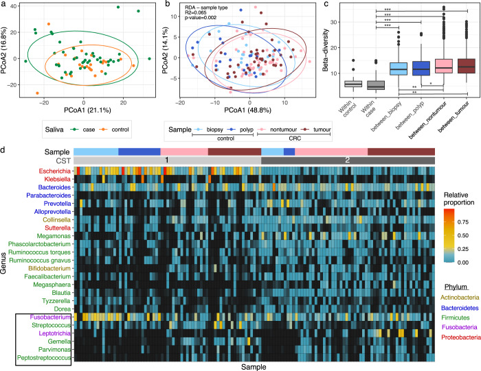
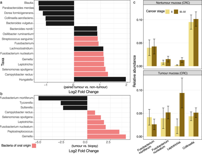
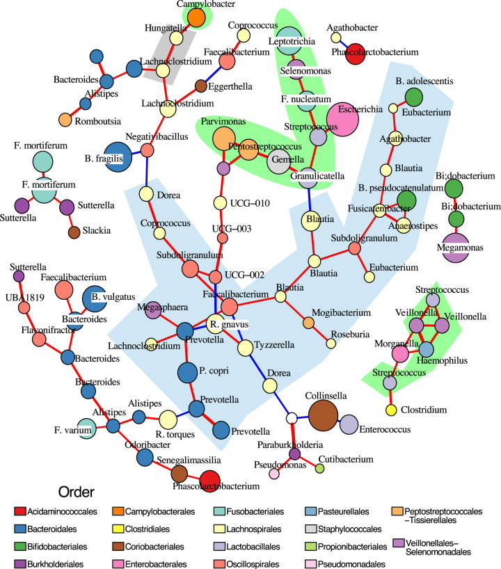
Perturbations in the gut microbiome have been associated with colorectal cancer (CRC), with the colonic overabundance of Fusobacterium nucleatum shown as the most consistent marker. Despite its significance in the promotion of CRC, genomic studies of Fusobacterium is limited. We enrolled 43 Vietnamese CRC patients and 25 participants with non-cancerous colorectal polyps to study the colonic microbiomes and genomic diversity of Fusobacterium in this population, using a combination of 16S rRNA gene profiling, anaerobic microbiology, and whole genome analysis. Oral bacteria, including F. nucleatum and Leptotrichia, were significantly more abundant in the tumour microbiomes. We obtained 53 Fusobacterium genomes, representing 26 strains, from the saliva, tumour and non-tumour tissues of six CRC patients. Isolates from the gut belonged to diverse F. nucleatum subspecies (nucleatum, animalis, vincentii, polymorphum) and a potential new subspecies of Fusobacterium periodonticum. The Fusobacterium population within each individual was distinct and in some cases diverse, with minimal intra-clonal variation. Phylogenetic analyses showed that within four individuals, tumour-associated Fusobacterium were clonal to those isolated from non-tumour tissues. Genes encoding major virulence factors (Fap2 and RadD) showed evidence of horizontal gene transfer. Our work provides a framework to understand the genomic diversity of Fusobacterium within the CRC patients, which can be exploited for the development of CRC diagnostic and therapeutic options targeting this oncobacterium.


Conflict of interest statement

The authors declare no competing interests.

Figures

Fig. 1
Fig. 1. The salivary and gut mucosal microbiomes of colorectal cancer patients.
Principal coordinate analyses (PCoA), conducted on phylogenetic-assisted isometric log-ratio (PhILR) transformed data, of (a) 66 salivary microbiomes, and (b) 129 gut mucosal microbiomes, with different CRC groups and sample types denoted by different colours (see Keys; biopsies and polyps collected from controls, nontumours and tumours collected from cases). c Boxplot showing the distribution of pairwise beta-diversity, calculated on PhILR transformed values, observed in each gut microbiome category. Bold central lines denote the median, the upper whisker extends from the 75th percentile to the highest value within the 1.5*interquartile range (IQR) of the hinge, the lower whisker extends from the 25th percentile to the lowest value within 1.5*IQR of the hinge. Data points beyond the end of the whiskers are outliers. Asterisk markings represent statistically significant differences between groups, as calculated by posthoc Tukey test (p-values ranging from >0.01 to ≤0.05 (*); from >1e−5 to ≤0.01 (**); ≤1e−5 (***)). d Heatmap displaying the proportional abundances of 24 most abundant genera (prevalence ≥15%, mean relative abundance ≥1%, and accounting for ~85% of the gut mucosal microbiome composition), with headers showing the samples’ community state type (CST): CST1 (light gray), CST2 (dark gray), and the corresponding sample type: biopsy (light blue), polyp (dark blue), nontumour (pink), tumour (dark red). Genera were coloured according to their classifications at Phylum level (see Keys). Genera in black box represent ones with probable origin from the oral cavity. The contributions of 24 genera listed here were summarized in Supplementary Table 3. Source data are provided as a Source Data file.
Fig. 2
Fig. 2. Bacterial taxa significantly abundant among the examined classes.
Taxa, or amplicon sequence variants (ASVs), were determined as significant and visualized in a and b if they were detected in at least two of the three tested approaches (ANCOMBC, DESeq2, corncob; adjusted p-value ≤ 0.05). a Log2 fold change of ASVs that differ between paired tumour and non-tumour mucosal microbiomes from case participants, using the full model ‘Patient + sample type’ (n = 86). b Log2 fold change of ASVs that differ between tumour and biopsy (control) mucosal microbiomes (n = 67). Log2 fold change was derived from ANCOMBC test output, and taxa of oral origin were coloured in pink. c Relative abundance of ASVs assigned as Fusobacterium mortiferum (n = 14), Fusobacterium nucleatum (n = 14), Leptotrichia (n = 16), and Collinsella (n = 14) in the tumour and nontumour mucosal microbiomes, stratified by cancer stages (III-IV vs. II). The error bar represents standard error of the mean relative abundance in each bar plot. Source data are provided as a Source Data file.
Fig. 3
Fig. 3. Correlation network of colorectal cancer gut mucosal microbiomes.
The network was constructed from 117 most representative ASVs sampled from 86 mucosal microbiomes, outlining significant interactions detected by both CCLasso (p value ≤ 0.01 and absolute correlation strength > 0.37) and SpiecEasi. Positive and negative interactions were coloured as red and blue lines respectively, with line weight proportional to correlation strength. The ASVs (nodes) were coloured based on taxonomic family (see Legend), with sizes proportional to their relative abundances. The light green shaded area entails ASVs identified as members of the human oral microbiome (comparison with expanded Human Oral Microbiome Database); the blue shaded area covers ASVs identified as gut anaerobic commensals (Lachnospirales, Bacteroidales, Bifidobacteriales, Oscillospirales); and gray shaded area covers other tumour-associated ASVs (as identified in Fig. 2a). Source data are provided as a Source Data file.
Fig. 4
Fig. 4. Maximum likelihood phylogenies of Fusobacterium isolates from this study.
a F. nucleatum species phylogeny constructed from the alignment of 516 core genes (89,900 variant sites; N = 57), using F. hwasookii clade as an outgroup. b F. periodonticum species phylogeny constructed from alignment of 863 core genes (106,738 variant sites, N = 26). Red circles at internal nodes denote bootstrap values ≥80. The associated metadata on the right describe the patient ID and clinical origin of isolates (where reference genomes were left blank), and the genomic presence of several virulence factors (fap2, fadA, fadA2, radD, fadA3, cbpF). Light blue shaded area covers isolates identified as novel F. periodonticum subspecies. The scale bars denote the estimated number of substitutions.

References

    1. Bray F, et al. Global cancer statistics 2018: GLOBOCAN estimates of incidence and mortality worldwide for 36 cancers in 185 countries. CA Cancer J. Clin. 2018;68:394–424. - PubMed
    1. Arnold M, et al. Global patterns and trends in colorectal cancer incidence and mortality. Gut. 2017;66:683–691. - PubMed
    1. Keum NN, Giovannucci E. Global burden of colorectal cancer: emerging trends, risk factors and prevention strategies. Nat. Rev. Gastroenterol. Hepatol. 2019;16:713–732. - PubMed
    1. Schirmer M, et al. Linking the human gut microbiome to inflammatory cytokine production capacity. Cell. 2016;167:1125–1136.e8. - PMC - PubMed
    1. Song M, Chan AT. Environmental factors, gut microbiota, and colorectal cancer prevention. Clin. Gastroenterol. Hepatol. 2019;17:275–289. - PMC - PubMed

Publication types

Substances