Review
. 2023 Feb;21(2):112-124.
doi: 10.1038/s41579-022-00809-7. Epub 2022 Oct 28.

SARS-CoV-2 variant evasion of monoclonal antibodies based on in vitro studies

Affiliations
Review

SARS-CoV-2 variant evasion of monoclonal antibodies based on in vitro studies

MacGregor Cox et al. Nat Rev Microbiol. 2023 Feb.

Abstract

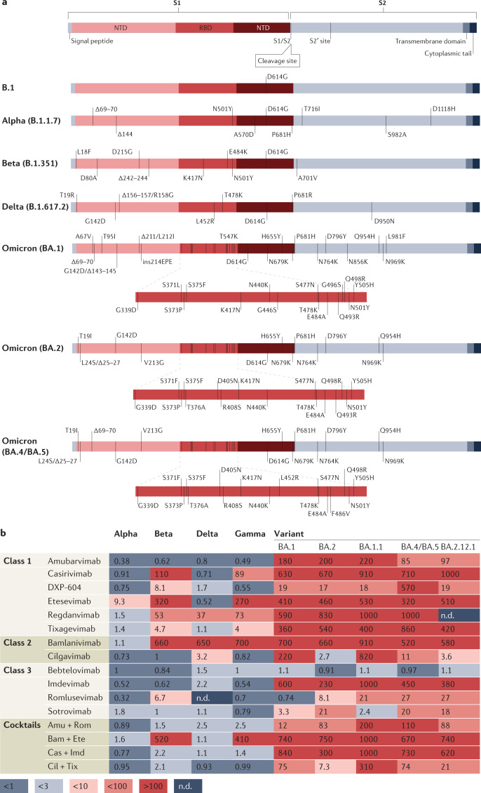
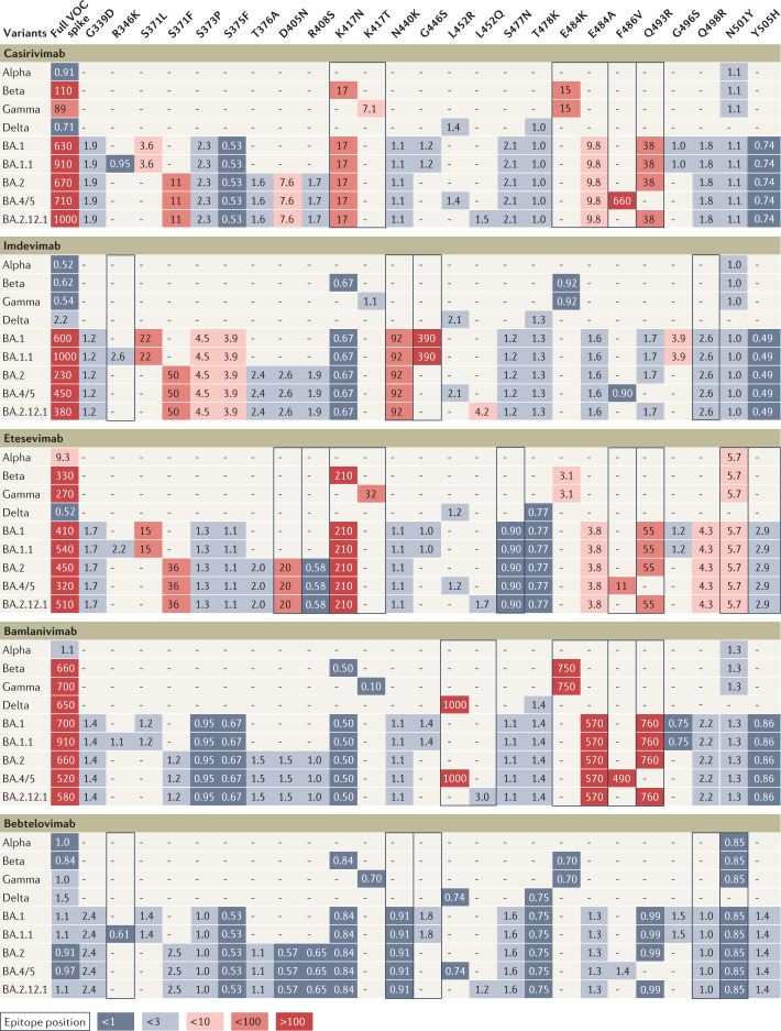
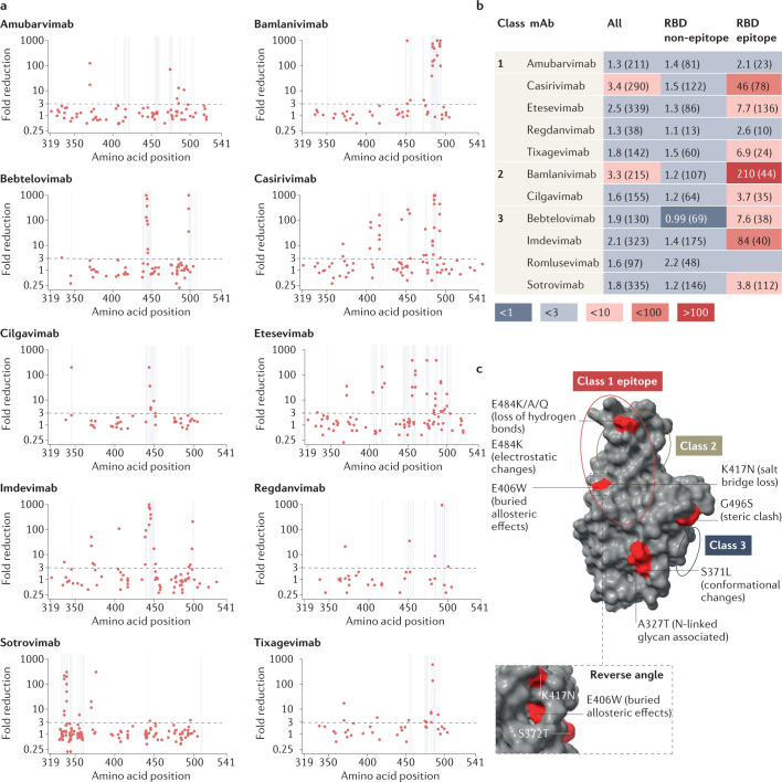
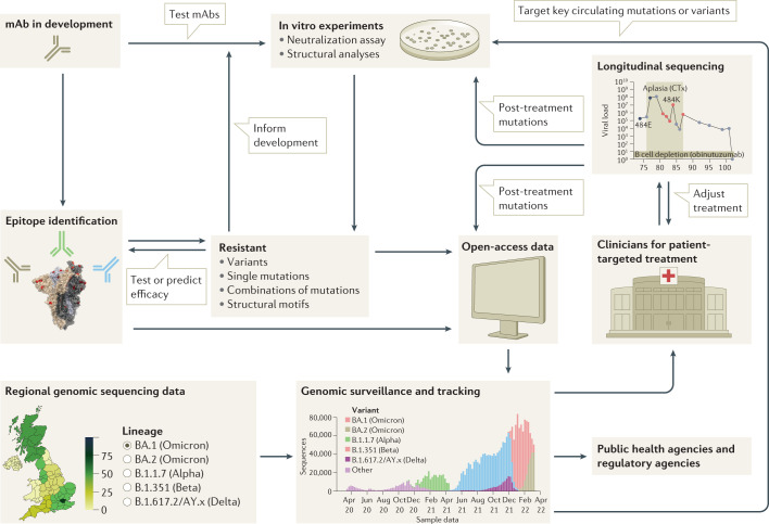
Monoclonal antibodies (mAbs) offer a treatment option for individuals with severe COVID-19 and are especially important in high-risk individuals where vaccination is not an option. Given the importance of understanding the evolution of resistance to mAbs by SARS-CoV-2, we reviewed the available in vitro neutralization data for mAbs against live variants and viral constructs containing spike mutations of interest. Unfortunately, evasion of mAb-induced protection is being reported with new SARS-CoV-2 variants. The magnitude of neutralization reduction varied greatly among mAb-variant pairs. For example, sotrovimab retained its neutralization capacity against Omicron BA.1 but showed reduced efficacy against BA.2, BA.4 and BA.5, and BA.2.12.1. At present, only bebtelovimab has been reported to retain its efficacy against all SARS-CoV-2 variants considered here. Resistance to mAb neutralization was dominated by the action of epitope single amino acid substitutions in the spike protein. Although not all observed epitope mutations result in increased mAb evasion, amino acid substitutions at non-epitope positions and combinations of mutations also contribute to evasion of neutralization. This Review highlights the implications for the rational design of viral genomic surveillance and factors to consider for the development of novel mAb therapies.


Conflict of interest statement

R.K.G. has received honoraria for educational activities from Moderna, GSK and Janssen. The other authors declare no competing interests.

Figures

Fig. 1
Fig. 1. SARS-CoV-2 variant of concern evasion of neutralization by monoclonal antibodies.
a, Spike mutational profiles for each variant of concern (VOC) with the receptor binding domain (RBD) also shown for BA.1, BA.2, and BA.4 and BA.5. b, Geometric mean fold reduction in neutralization (mFRN) values of monoclonal antibodies (mAbs) against VOCs relative to reference or control variants. Means are calculated from published studies reporting neutralization data on clinically approved mAbs against VOCs. Data for the associated single studies are shown in Supplementary Fig. 2. Full data sets are available in Supplementary Data File 1, and confidence statistics in Supplementary Data File 2. Colours depict the strength of resistance: dark red, strong (mFRN > 100); red, moderate (mFRN = 10–100); light red, mild (mFRN = 3–10); light grey, no resistance (mFRN = 1–3); dark grey, increased sensitivity (mFRN < 1). All mFRN values are given to two significant figures. Amu + Rom, amubarvimab + romlusevimab; Bam + Ete, bamlanivimab + etesevimab; Cas + Imd, casirivimab + imdevimab; Cil + Tix, cilgavimab + tixagevimab; n.d., no neutralization data reported for the antibody–variant pair at the time of writing; NTD, amino-terminal domain.
Fig. 2
Fig. 2. Influence of individual spike mutations of interest on monoclonal antibody neutralization activity compared with antibody resistance of variants.
Heat map depicting fold change in monoclonal antibody (mAb) neutralization for variants of concern (VOCs) (left-most column) and individual mutations (other columns). Each row contains data for the full spike profile of a given VOC, as well as each definitive VOC mutation individually on a wild type background. Comparison of fold change values across a row indicates which mutations are responsible for any resistance shown by the full VOC. Values show geometric mean fold reduction in neutralization (mFRN). Boxes indicate epitope positions (see Supplementary Data File 3). Colours depict the strength of resistance: dark red, strong (mFRN > 100); red, moderate (mFRN = 10–100); light red, mild (mFRN = 3–10); light grey, no resistance (mFRN = 1–3); dark grey, increased sensitivity (mFRN < 1). ‘–’ indicates that the mutation is not present in the variant. All defining mutations at receptor binding domain (RBD) positions in VOCs are included. Supplementary Fig. 3 presents data for other mAbs for which less comprehensive data have been collected. The RBD is defined here as spike amino acid positions 319–541 (ref.).
Fig. 3
Fig. 3. Assessment of monoclonal antibody resistance by mutations of interest occurring at epitope and non-epitope sites.
a, Geometric mean fold reduction in neutralization (mFRN) data for each monoclonal antibody (mAb) against viral constructs containing single mutations in the spike receptor binding domain (RBD) (positions 319–541). Epitope positions (see Supplementary Data File 3) indicated by vertical grey lines. Dashed line shows the mFRN = 3 threshold. Alternative substitutions at the same amino acid position are shown as separate points at the same x coordinate, for example E484K and E484Q are shown as two different points at the 484 position on the x axis. Most dots above the threshold coincide with the grey lines, indicating that mutations conferring resistance to neutralization tend to occur at epitope positions. However, some non-epitope mutations also confer resistance as shown by the dots above the threshold but not on grey lines. b, Pooled mFRN comparisons between epitope and non-epitope mutations. Values shown are mFRN for either all amino acid positions, all amino acid positions in the epitope of the mAb, or all amino acid positions in the RBD but not in the epitope of the mAb. The higher mFRN values of epitope positions indicate the increased contribution of mutations in the mAb epitope to the resistance of neutralization. Number in brackets indicates the number of assays contributing to each geometric mean value. Epitope unknown for romlusevimab. Colours depict the strength of resistance: dark red, strong (mFRN > 100); red, moderate (mFRN = 10–100); light red, mild (mFRN = 3–10); light grey, no resistance (mFRN = 1–3); dark grey, increased sensitivity (mFRN < 1). c, Isolated Omicron spike RBD structure [PDB:7TGW]. Epitope regions for class 1, 2 and 3 mAbs are circled. Epitope and non-epitope mutations exemplifying mechanisms of mAb evasion are labelled: S371L, conformational changes and N-linked glycosylation; A372T, N-linked glycosylation; E406W, conformational changes in the epitope; K417N, abolished salt bridges between mAb and RBD; E484K/E484Q/E484A, loss of hydrogen bonds with mAb; E484K, changes to electrostatic interactions; G496S, steric clash. Single mutant mFRN data across the full spike protein are shown in Supplementary Fig. 6a and pooled mFRN comparisons between epitope, non-epitope mutations, epitope proximal and RBD positions in Supplementary Fig. 6b.
Fig. 4
Fig. 4. Framework for the rational design of viral genomic surveillance for the development of efficient monoclonal antibody therapies.
A successful framework will integrate knowledge of the dynamics of monoclonal antibody (mAb) resistance by SARS-CoV-2 variants, including the central role played by epitope mutations, epistatic effects and evolutionary dynamics. mAb development must be supported by epitope identification, in vitro studies and the monitoring of resistance mutations. mAbs in early development may be ruled out if they target an epitope that contains mutations shown to cause resistance in vitro. Alternatively, low frequency of, or evolutionary barriers to, mutations in a given region may focus the development of mAbs that target that region. Resistance monitoring is a multifaceted process, spanning individual and combinatorial mutational effects in vitro, as well as genomic surveillance of circulating strains and longitudinal clinical studies that track genetic changes during treatment. Combining these different approaches will allow the development of mAbs highly effective against currently circulating strains, and robust to resistance by future variants.

Similar articles

Cited by

References

    1. World Health Organization. WHO COVID-19 dashboard. World Health Organizationhttps://covid19.who.int/ (2022).
    1. Uddin M, et al. SARS-CoV-2/COVID-19: viral genomics, epidemiology, vaccines, and therapeutic interventions. Viruses. 2020;12:526. doi: 10.3390/v12050526. - DOI - PMC - PubMed
    1. Drożdżal S, et al. An update on drugs with therapeutic potential for SARS-CoV-2 (COVID-19) treatment. Drug Resist. Updat. 2021;59:100794. doi: 10.1016/j.drup.2021.100794. - DOI - PMC - PubMed
    1. Zhou L, Ayeh SK, Chidambaram V, Karakousis PC. Modes of transmission of SARS-CoV-2 and evidence for preventive behavioral interventions. BMC Infect. Dis. 2021;21:496. doi: 10.1186/s12879-021-06222-4. - DOI - PMC - PubMed
    1. Luo L, Luo T, Du M, Mei H, Hu Y. Efficacy and safety of tocilizumab in hospitalized COVID-19 patients: a systematic review and meta-analysis. J. Infect. 2022;84:418–467. doi: 10.1016/j.jinf.2021.11.013. - DOI - PMC - PubMed

Publication types

Supplementary concepts