. 2022 Oct 28;12(1):177.
doi: 10.1186/s13578-022-00915-2.

Wasteosomes (corpora amylacea) of human brain can be phagocytosed and digested by macrophages

Affiliations

Wasteosomes (corpora amylacea) of human brain can be phagocytosed and digested by macrophages

Marta Riba et al. Cell Biosci. .

Abstract

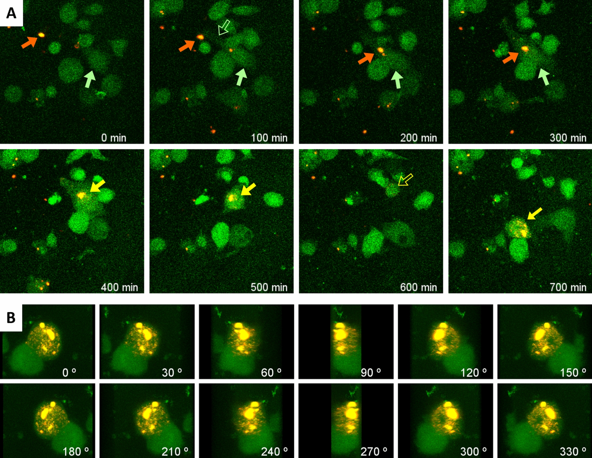
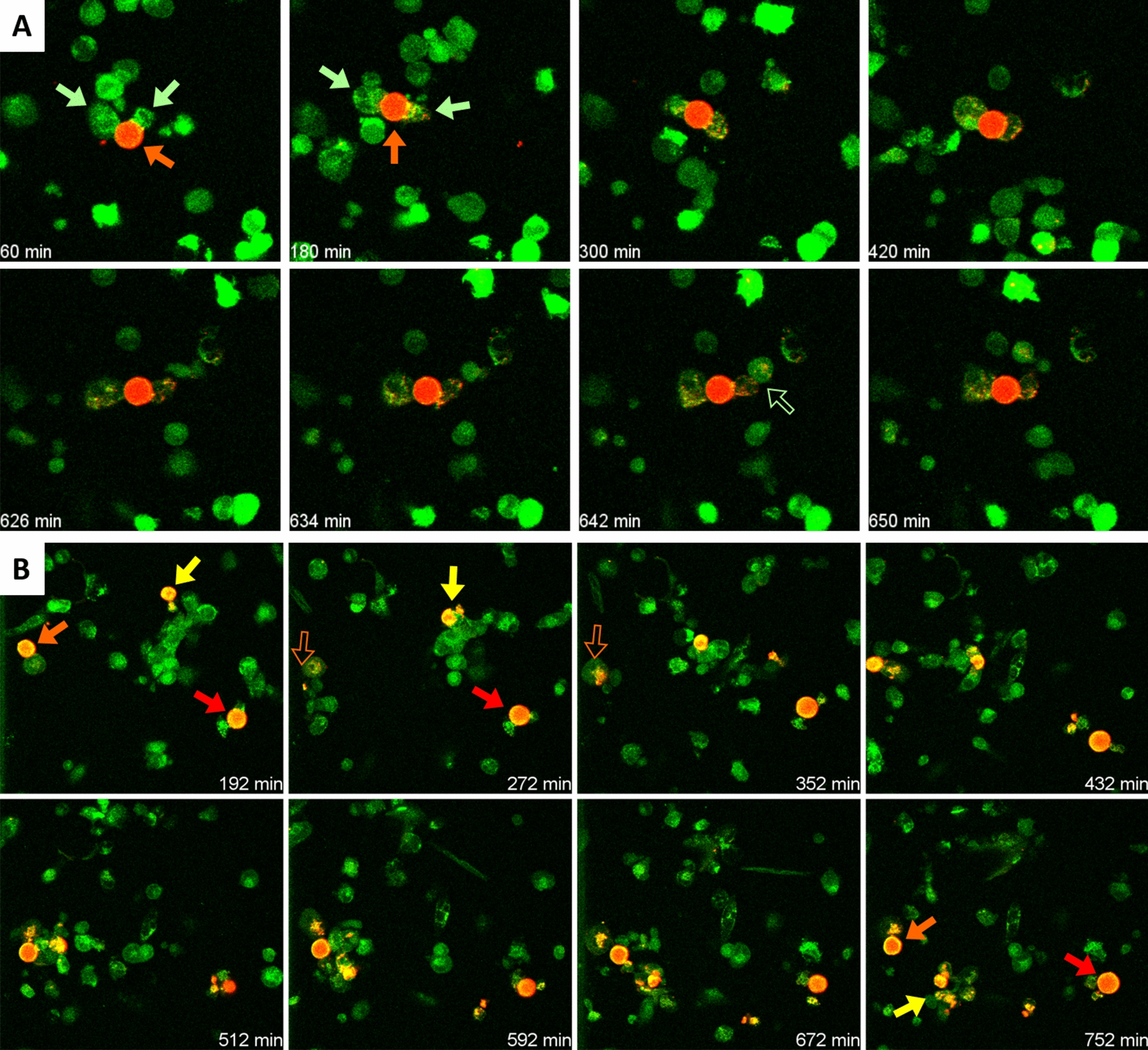
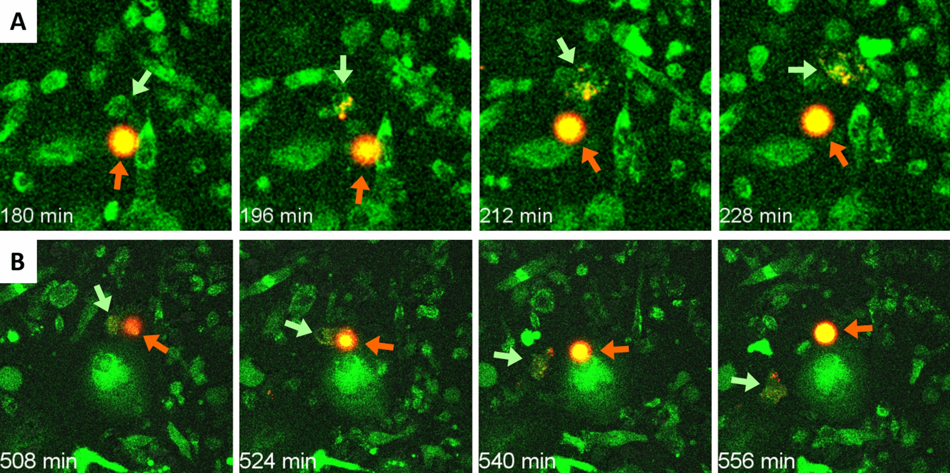
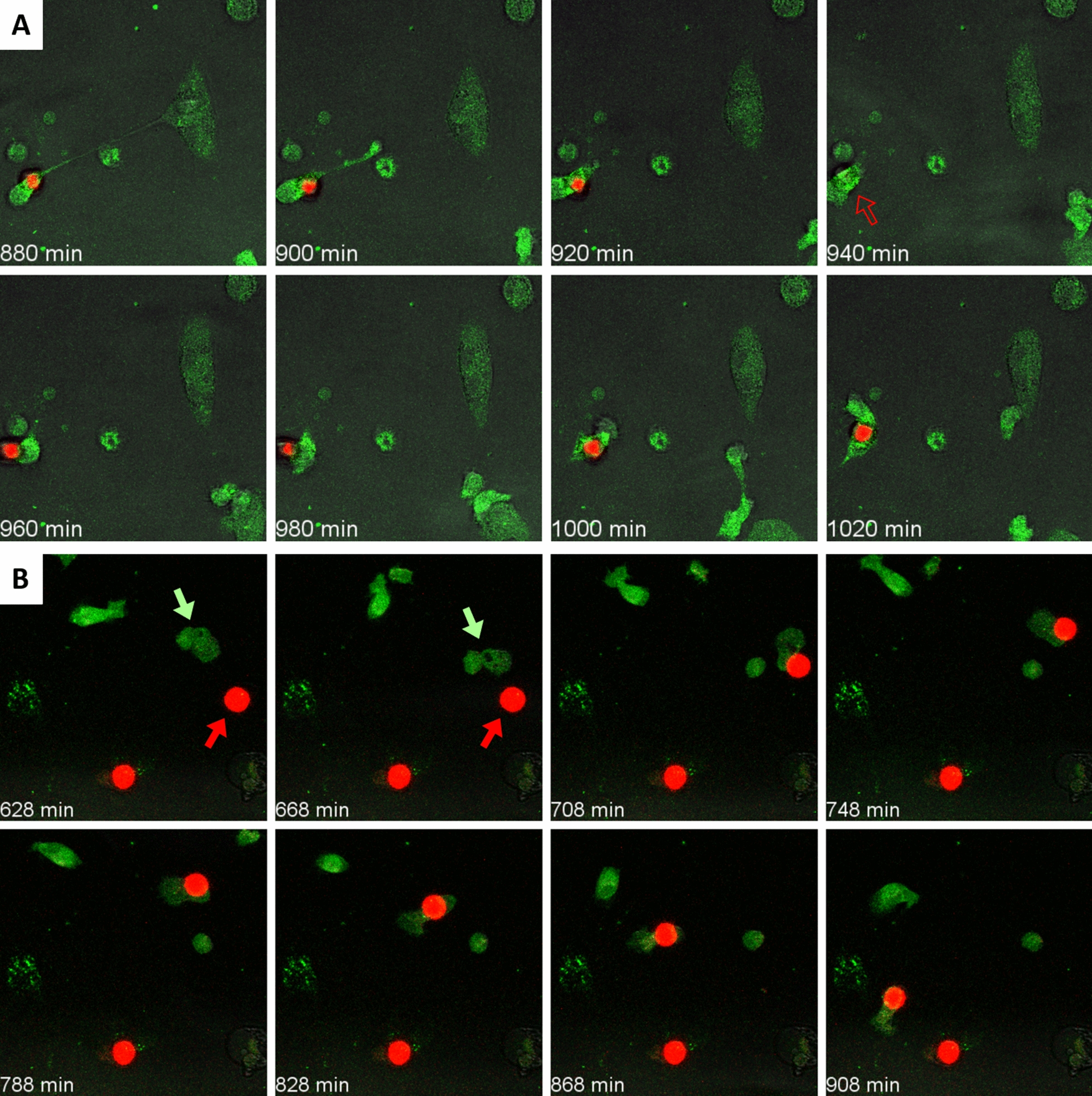
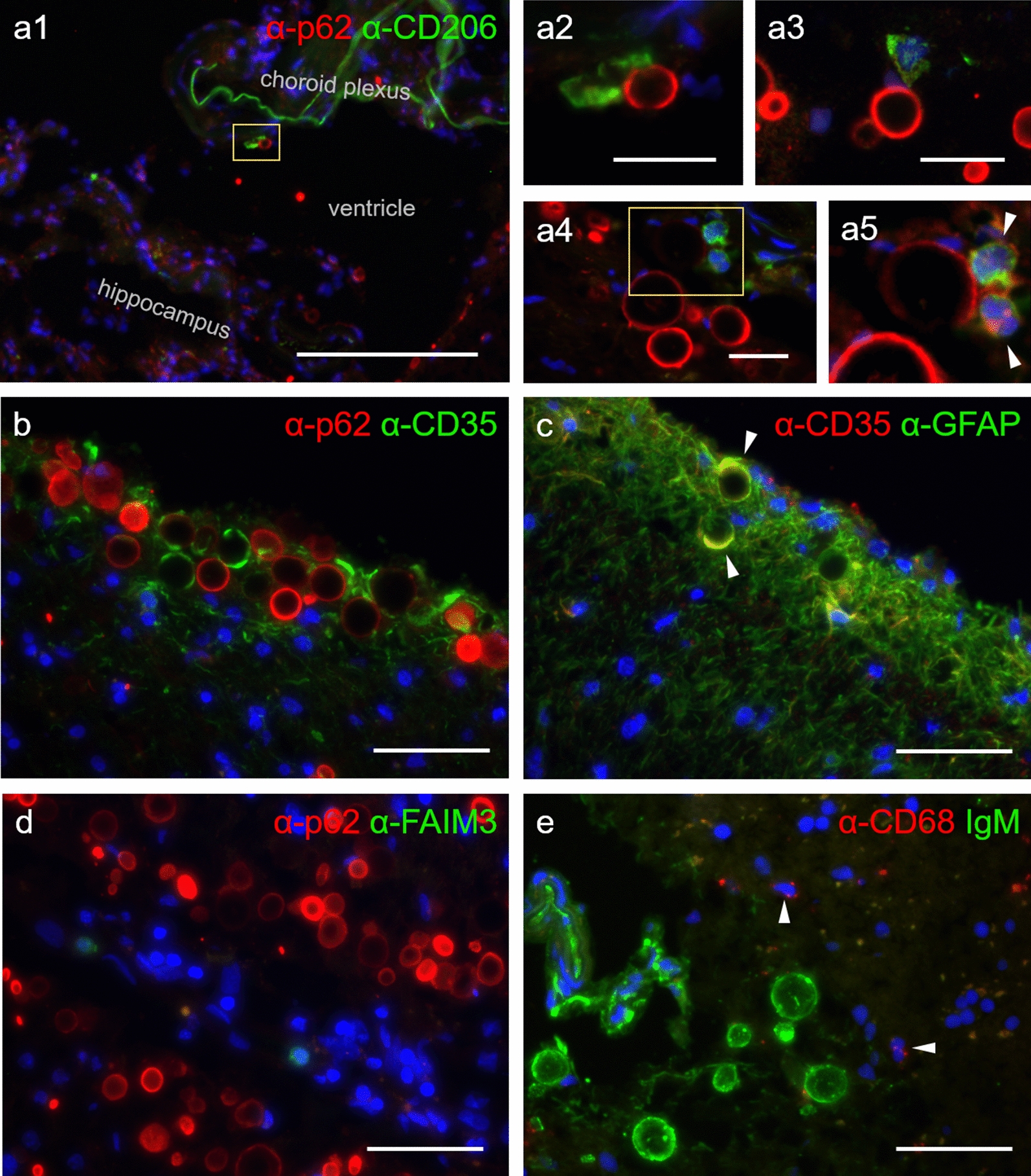
Background: Corpora amylacea of human brain, recently renamed as wasteosomes, are granular structures that appear during aging and also accumulate in specific areas of the brain in neurodegenerative conditions. Acting as waste containers, wasteosomes are formed by polyglucosan aggregates that entrap and isolate toxic and waste substances of different origins. They are expelled from the brain to the cerebrospinal fluid (CSF), and can be phagocytosed by macrophages. In the present study, we analyze the phagocytosis of wasteosomes and the mechanisms involved in this process. Accordingly, we purified wasteosomes from post-mortem extracted human CSF and incubated them with THP-1 macrophages. Immunofluorescence staining and time-lapse recording techniques were performed to evaluate the phagocytosis. We also immunostained human hippocampal sections to study possible interactions between wasteosomes and macrophages at central nervous system interfaces.

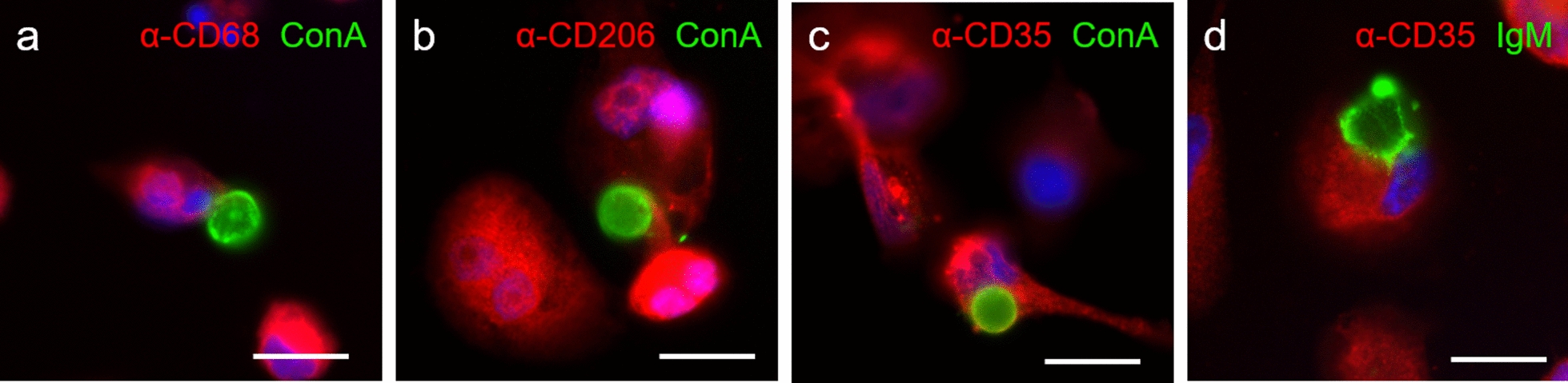
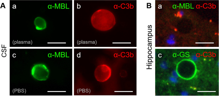
Results: We observed that the wasteosomes obtained from post-mortem extracted CSF are opsonized by MBL and the C3b complement protein. Moreover, we observed that CD206 and CD35 receptors may be involved in the phagocytosis of these wasteosomes by THP-1 macrophages. Once phagocytosed, wasteosomes become degraded and some of the resulting fractions can be exposed on the surface of macrophages and interchanged between different macrophages. However, brain tissue studies show that, in physiological conditions, CD206 but not CD35 receptors may be involved in the phagocytosis of wasteosomes.

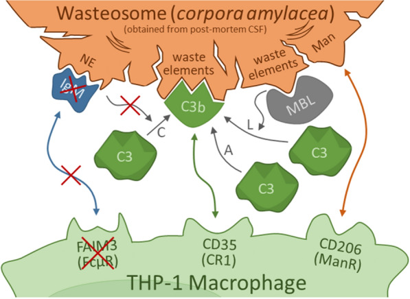
Conclusions: The present study indicates that macrophages have the machinery required to process and degrade wasteosomes, and that macrophages can interact in different ways with wasteosomes. In physiological conditions, the main mechanism involve CD206 receptors and M2 macrophages, which trigger the phagocytosis of wasteosomes without inducing inflammatory responses, thus avoiding tissue damage. However, altered wasteosomes like those obtained from post-mortem extracted CSF, which may exhibit waste elements, become opsonized by MBL and C3b, and so CD35 receptors constitute another possible mechanism of phagocytosis, leading in this case to inflammatory responses.

Keywords: Brain; C3b; CD206; CD35; Corpora amylacea; IgM; Natural immunity; Phagocytosis; Wasteosome.


Conflict of interest statement

The authors declare that they have no competing interests.

Figures

Fig. 1
Fig. 1
A Sequence of images from a time-lapse recording showing how a THP-1 macrophage (green arrow) extends a lamellipodium (empty green arrow) to a wasteosome opsonized with ConA (red arrow) and pulls it towards the body of the macrophage, triggering the engulfment of the wasteosome. The phagocytosed wasteosome become later digested and fragmented (yellow arrows). Empty yellow arrow: wasteosome is out of the focus plane. See video for details. At 1202 min, confocal images were taken and the 3D reconstruction was made. B Sequence of images showing the 360° rotation of the 3D reconstruction. Images permit to observe the location of the remains of the wasteosome (red and yellow) at the macrophage. Some dots of red or yellow fluorescence appear on the surface of the macrophage, suggesting antigen presentation. The big green spot in the lower region corresponds to a macrophage that is in contact with the one that has phagocytosed and digested the wasteosome
Fig. 2
Fig. 2
A Sequence of images from a time-lapse recording showing two THP-1 macrophages (green arrows) eroding a wasteosome opsonized with ConA (red arrow). Some spots of red fluorescence become incorporated into the macrophages. In some cases, the fluorescence is transferred from one macrophage to another one (empty green arrow). See video for details. B Sequence of images showing different macrophages interacting with three different wasteosomes (arrows). One of the wasteosomes (yellow arrow) become digested and fragmented. Empty arrows indicate that the wasteosome is out of the focus plane
Fig. 3
Fig. 3
A Sequence of images showing a THP-1 macrophage (green arrow) eroding a wasteosome opsonized with AF555-NHS (red arrow). Note that the spots of red fluorescence, corresponding to stained proteins, become incorporated into the macrophage. A similar process can be observed in B. See the corresponding videos for details
Fig. 4
Fig. 4
A Sequence of images showing a THP-1 macrophage (green) contacting a wasteosome stained with PAS (red). Initially, a lamellipodium from a distant macrophage making contact with the wasteosome can be observed. Thereafter, the macrophage shrinks over the wasteosome and both structures displace together. However, unlike in the previous sets of experiments, the digestion of the PAS stained wasteosomes could not be observed in most of the experiments. B In only a few cases, as the one shown here, small amounts of fluorescence were observed to detach from the wasteosomes and spread inside the macrophages. See videos for details
Fig. 5
Fig. 5
THP-1 macrophages which make contact with wasteosomes are CD68 + , CD206 + and CD35 + . a A CD68 + THP-1 macrophage (red) in contact with a wasteosome stained with ConA (green). b A CD206 + THP-1 macrophage (red) attached to a wasteosome (ConA, green). c A CD35 + THP-1 macrophage (red) encircling a wasteosome stained with ConA (green). d A CD35 + THP-1 macrophage (red) encircling a wasteosome immunostained with IgM (green). Nuclei are stained with Hoechst (blue). Scale bar: 25 µm
Fig. 6
Fig. 6
A Wasteosomes from CSF have opsonins on their surface. a wasteosomes purified from CSF and incubated with human plasma become stained with anti-MBL (green). b wasteosomes from CSF and incubated with human plasma become immunostained with anti-C3b (red). c wasteosomes purified from CSF and incubated with PBS instead of human plasma also become immunostained with anti-MBL (green). d wasteosomes purified from CSF and incubated with PBS instead of human plasma also become immunostained with anti-C3b (red). B Hippocampal wasteosomes do not have the opsonins on their surface. a When the hippocampal tissue is double-immunostained with anti-MBL (green) and anti-C3b (red), the wasteosomes do not become immunostained and are observed as a black circle. In this case, one wasteosome can be observed in the center of the image. b When the hippocampal tissue is immunostained with anti-GS and anti-C3b, wasteosomes become stained with anti-GS (green) but not by anti-C3b (red). Scale bars: 25 µm
Fig. 7
Fig. 7
Some macrophages at central nervous system interfaces interact with wasteosomes. a1, a2, a3, a4 and a5 wasteosomes from human hippocampal sections immunostained with anti-p62 (red) and interface macrophages immunostained with anti-CD206 (green). a1 A choroid plexus macrophage in contact with a wasteosome released from the brain tissue to the ventricular CSF. a2 inset of a1, where the macrophage attached to the wasteosome is magnified. a3 and a4 wasteosomes released from hippocampus to the subarachnoid space in contact with meningeal macrophages. a5 inset of a4, where the red staining is digitally intensified to evidence the presence of a wasteosome, in this case surrounded by two macrophages (white arrowheads). b within the brain parenchyma, wasteosomes become immunostained with anti-p62 (red) and are surrounded by CD35 + cells (green). c CD35 staining (red) colocalize with GFAP staining (green, white arrowheads), indicating that cells that surround wasteosomes are CD35 + astrocytes. d wasteosomes from human hippocampus immunostained with anti-p62. FAIM3 positive cells are not found in the hippocampal sections (green). e wasteosomes from human hippocampal tissue immunostained with IgMs (green) are not contacted by CD68 + cells (red, marked with white arrowheads). For their localization, these CD68 + cells are presumably microglial cells, although possible infiltrating macrophages cannot be discarded. In any case, and as expected, microglia or macrophages did not reach the wasteosomes within the brain parenchyma since in this region wasteosomes are intracellular astrocytic structures. Scale bar in a1: 200 µm; scale bars in a2, a3 and a4: 25 µm; other scale bars: 50 μm. Hoechst (blue) was used for nuclear staining
Fig. 8
Fig. 8
Integrative scheme of the potential mechanisms involved in the phagocytosis of wasteosomes obtained from post-mortem extracted CSF by THP-1 macrophages.The absence of FAIM3 in THP-1 macrophages and the absence of IgM opsonizing the CSF wasteosomes (binding to their NEs) indicate that the phagocytosis by THP-1 macrophages does not occur through the IgM-FAIM3 interaction. However, phagocytosis could occur through CD206 and CD35 receptors, which are both present in THP-1 macrophages. In the first case, the CD206 receptor can interact with mannose or other carbohydrates located in the wasteosomes. In the second one, the CD35 receptor can recognize the C3b that opsonizes wasteosomes once they arrive at the CSF. The opsonization with C3b in the CSF can be produced through the lectin pathway (L), triggered by the binding of MBL with the wasteosomes, and through the alternative pathway (A), triggered by the contact between C3 and wasteosomes, but not through the complement pathway (C), because IgM do not opsonize wasteosomes in the CSF. Note that wasteosomes are obtained from post-mortem extracted CSF and are altered wasteosomes that may show waste elements. At the central nervous system interfaces, in which waste elements are enclosed into the polyglucosan structure, the wasteosomes are not opsonized by MBL or C3b, and they interact with CD206 + macrophages but not with CD35 + or FAIM3 + macrophages. See text for details

Similar articles

Cited by

References

    1. Sakai M, Austin J, Witmer F, Trueb L. Studies of corpora amylacea. Arch Neurol. 1969;21:526–544. doi: 10.1001/archneur.1969.00480170098011. - DOI - PubMed
    1. Sbarbati A, Carner M, Colletti V, Osculati F. Extrusion of corpora amylacea from the marginal glia at the vestibular root entry zone. J Neuropathol Exp Neurol. 1996;55:196–201. doi: 10.1097/00005072-199602000-00008. - DOI - PubMed
    1. Cavanagh JB. Corpora-Amylacea and the family of polyglucosan diseases. Brain Res Rev. 1999;29:265–295. doi: 10.1016/S0165-0173(99)00003-X. - DOI - PubMed
    1. Singhrao SK, Neal JW, Piddlesden SJ, Newman GR. New immunocytochemical evidence for a neuronal/oligodendroglial origin for corpora amylacea. Neuropathol Appl Neurobiol. 1994;20:66–73. doi: 10.1111/j.1365-2990.1994.tb00958.x. - DOI - PubMed
    1. Radhakrishnan A, Radhakrishnan K, Radhakrishnan VV, Mary PR, Kesavadas C, Alexander A, et al. Corpora amylacea in mesial temporal lobe epilepsy: clinico-pathological correlations. Epilepsy Res. 2007;74:81–90. doi: 10.1016/j.eplepsyres.2007.01.003. - DOI - PubMed

LinkOut - more resources