

New insight into the role of lipid metabolism-related proteins in rheumatic heart valve disease
- PMID: 36307855
- PMCID: PMC9615153
- DOI: 10.1186/s12944-022-01722-x
New insight into the role of lipid metabolism-related proteins in rheumatic heart valve disease
Abstract
Purpose: The aim of this study was to determine the expression of lipid metabolism-related proteins in rheumatic heart valve disease (RHVD).
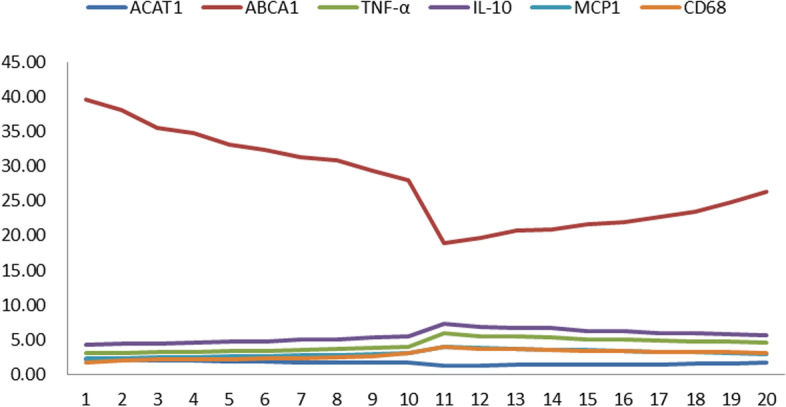
Methods: This retrospective study involved a total of 20 cases of moderate or severe rheumatic mitral valve stenosis and 4 cases of mitral regurgitation due to secondary causes from September 2018 to September 2021. The patients enrolled included 12 males and 12 females who underwent surgical excision of the mitral valve at the cardiac surgery department of Hainan General Hospital. The samples of mitral valve were collected during surgery treatment as the study group, and mitral valves collected from patients with ischemic heart disease were allocated into the control group. Hematoxylin-eosin (HE), oil red staining and immunohistochemical (IHC) staining were conducted to compare the expression of lipid metabolism-related proteins (ATP-binding cassette transporter A1 and acyl-coenzyme A: cholesterol acyltransferase-1), and real-time polymerase chain reaction (RT-PCR) was applied to compare the mRNA levels of ABCA1, ACAT1, and the inflammatory cytokines TNF-α, IL-10, and MCP-1.
Results: In general, the rheumatic mitral valve showed leaflet thickening along with border adhesions and visible yellow fats. Oil red O staining also revealed the abovementioned results as well as fat cells. Both ABCA1 and ACAT1 were expressed in the rheumatic mitral valve via IHC, whereas only ACAT1 showed a faint level of expression in the ischemic mitral valve with no expression of ABCA1. In addition, compared with the ischemic mitral valve, RT-PCT showed increased mRNA expression levels of ABCA1, ACAT1, and the inflammatory cytokines TNF-α, IL-10, and MCP-1 (P < 0.05). After dividing the RMs into two groups for RT-PCR, we found that the higher the expression of ABCA1 and ACAT1 was, the lower the relative expression of inflammatory factors.
Conclusion: This study showed that adipose tissue, adipose cells, and lipid transport-related proteins were expressed strongly in the rheumatic mitral valve, suggesting that adipose tissue formation might be one of the important pathways in the pathology of rheumatic heart disease. In addition, adipose tissue and adipocytes were also involved in the inflammatory process. These data provide new insight into pathological mechanisms in rheumatic heart disease.
Keywords: Hematoxylin–eosin staining; Immunohistochemical staining; Lipid metabolism; Rheumatic mitral valve disease.
© 2022. The Author(s).
Conflict of interest statement
The authors declare that they have no competing interests.
Figures


Similar articles
-
A new perspective: Fat tissue and adipokines in rheumatic heart valves.J Card Surg. 2022 Dec;37(12):4991-4998. doi: 10.1111/jocs.17216. Epub 2022 Nov 24. J Card Surg. 2022. PMID: 36423241
-
IL-10 and ET-1 as biomarkers of rheumatic valve disease.Rev Bras Cir Cardiovasc. 2014 Jan-Mar;29(1):25-30. doi: 10.5935/1678-9741.20140007. Rev Bras Cir Cardiovasc. 2014. PMID: 24896159 Free PMC article.
-
Analysis of the Correlation Between the Expression of T-Helper Type 17 Cell-Related Cytokines and Valve Damage in Rheumatic Heart Disease.Cureus. 2024 Oct 31;16(10):e72759. doi: 10.7759/cureus.72759. eCollection 2024 Oct. Cureus. 2024. PMID: 39618620 Free PMC article.
-
Rheumatic mitral valve disease: current surgical status.Prog Cardiovasc Dis. 2009 May-Jun;51(6):478-81. doi: 10.1016/j.pcad.2008.08.008. Prog Cardiovasc Dis. 2009. PMID: 19410681 Review.
-
Current status of surgery for rheumatic carditis in children.Ann Thorac Surg. 2004 Oct;78(4):1403-8. doi: 10.1016/j.athoracsur.2004.04.079. Ann Thorac Surg. 2004. PMID: 15464505 Review.
Cited by
-
Causal effects of inflammatory cytokines on cardiovascular diseases: Insights from genetic evidence.Heliyon. 2024 Jul 30;10(15):e35447. doi: 10.1016/j.heliyon.2024.e35447. eCollection 2024 Aug 15. Heliyon. 2024. PMID: 39165962 Free PMC article.
References
MeSH terms
Substances
LinkOut - more resources
Full Text Sources
Medical
Miscellaneous
