

Differentiating tracheobronchial involvement in granulomatosis with polyangiitis and relapsing polychondritis on chest CT: a cohort study
- PMID: 36307863
- PMCID: PMC9615207
- DOI: 10.1186/s13075-022-02935-2
Differentiating tracheobronchial involvement in granulomatosis with polyangiitis and relapsing polychondritis on chest CT: a cohort study
Abstract
Background: In patients with tracheobronchial involvement, the differential diagnosis between granulomatosis with polyangiitis (GPA) and relapsing polychondritis (RP) can be challenging. The aim of this study was to describe the characteristics of airway abnormalities on chest computed tomography (CT) in patients with GPA or RP and to determine whether specific imaging criteria could be used to differentiate them.
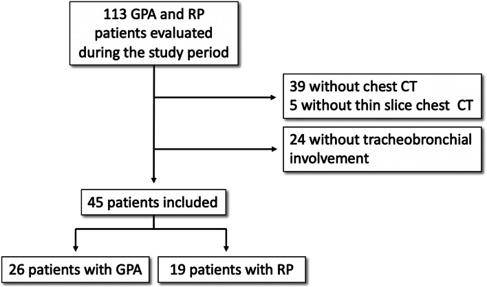
Methods: GPA and RP patients with tracheobronchial involvement referred to a national referral center from 2008 to 2020 were evaluated. Their chest CT images were reviewed by two radiologists who were blinded to the final diagnosis in order to analyze the characteristics of airway involvement. The association between imaging features and a diagnosis of GPA rather than RP was analyzed using a generalized linear regression model.
Results: Chest CTs from 26 GPA and 19 RP patients were analyzed. Involvement of the subglottic trachea (odds ratio for GPA=28.56 [95% CI: 3.17; 847.63]; P=0.001) and extensive airway involvement (odds ratio for GPA=0.02 [95% CI: 0.00; 0.43]; P=0.008) were the two independent CT features that differentiated GPA from RP in multivariate analysis. Tracheal thickening sparing the posterior membrane was significantly associated to RP (odds ratio for GPA=0.09 [95% CI: 0.02; 0.39]; P=0.003) but only in the univariate analysis and suffered from only moderate interobserver agreement (kappa=0.55). Tracheal calcifications were also associated with RP only in the univariate analysis (odds ratio for GPA=0.21 [95% CI: 0.05; 0.78]; P=0.045).
Conclusion: The presence of subglottic involvement and diffuse airway involvement are the two most relevant criteria in differentiating between GPA and RP on chest CT. Although generally considered to be a highly suggestive sign of RP, posterior tracheal membrane sparing is a nonspecific and an overly subjective sign.
Keywords: Granulomatosis with polyangiitis; Multidetector computed tomography; Relapsing polychondritis; Respiratory tract diseases; Trachea.
© 2022. The Author(s).
Conflict of interest statement
XP has been an investigator in academic studies of ANCA-associated vasculitis for which rituximab was provided by Roche Pharma. MPR reports personal fees from Boehringer Ingelheim, Bracco, Chiesi, and GE Healthcare; travel fees from Guerbet; and a research grant from the French Ministry of Health and the French Cancer Institute, outside the submitted work. G. C. reports personal fees from Chiesi and Gleamer outside the submitted work. None declared: C. J., I. S., Em. C., B. T., L. M., Eg. C., and S. M.
Figures


Similar articles
-
Presentation, Diagnosis, and Management of Subglottic and Tracheal Stenosis During Systemic Inflammatory Diseases.Chest. 2022 Jan;161(1):257-265. doi: 10.1016/j.chest.2021.07.037. Epub 2021 Jul 26. Chest. 2022. PMID: 34324839
-
[Characteristics of airway involvement in relapsing polychondritis].Zhonghua Yi Xue Za Zhi. 2006 Apr 18;86(15):1048-51. Zhonghua Yi Xue Za Zhi. 2006. PMID: 16784709 Chinese.
-
Subglottic stenosis and endobronchial disease in granulomatosis with polyangiitis.Rheumatology (Oxford). 2019 Dec 1;58(12):2203-2211. doi: 10.1093/rheumatology/kez217. Rheumatology (Oxford). 2019. PMID: 31199488 Free PMC article.
-
Tracheobronchial involvement of relapsing polychondritis.Autoimmun Rev. 2019 Sep;18(9):102353. doi: 10.1016/j.autrev.2019.102353. Epub 2019 Jul 16. Autoimmun Rev. 2019. PMID: 31323366 Review.
-
The presentation and management of granulomatosis with polyangiitis (Wegener's Granulomatosis) in the pediatric airway.Laryngoscope. 2017 Jan;127(1):233-240. doi: 10.1002/lary.26013. Epub 2016 Apr 26. Laryngoscope. 2017. PMID: 27113905 Review.
Cited by
-
Diagnostic Challenges and Management of Relapsing Polychondritis with Large-Airway Involvement: A Case Series and Literature Review.Life (Basel). 2024 Sep 21;14(9):1194. doi: 10.3390/life14091194. Life (Basel). 2024. PMID: 39337976 Free PMC article.
-
Relapsing polychondritis: tracheobronchial involvement and differential diagnoses.J Thorac Dis. 2025 Jan 24;17(1):461-475. doi: 10.21037/jtd-24-1603. Epub 2025 Jan 20. J Thorac Dis. 2025. PMID: 39975747 Free PMC article. Review.
-
Relapsing polychondritis: clinical updates and new differential diagnoses.Nat Rev Rheumatol. 2024 Jun;20(6):347-360. doi: 10.1038/s41584-024-01113-9. Epub 2024 May 2. Nat Rev Rheumatol. 2024. PMID: 38698240 Review.
References
MeSH terms
LinkOut - more resources
Full Text Sources
Medical
Research Materials
