

Effect of Propofol and Etomidate on the Proliferation, Cell-cycle Distribution, Apoptosis and Necrosis of Pancreatic Tumour Cells
- PMID: 36309382
- PMCID: PMC9677759
- DOI: 10.21873/invivo.13008
Effect of Propofol and Etomidate on the Proliferation, Cell-cycle Distribution, Apoptosis and Necrosis of Pancreatic Tumour Cells
Abstract
Background/aim: The influence of surgical interventions and anaesthesiological procedures on tumour progression was investigated as early as the 1920s. In current cancer management, the perioperative phase is increasingly being considered a vulnerable period with an increased risk of tumour cell dissemination due to medication, surgical manipulation, and immunosuppression. The extent to which narcotics administered in the perioperative setting influence the oncological outcomes of patients with pancreatic cancer is still unclear.
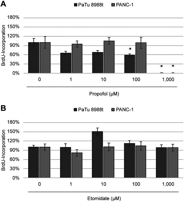
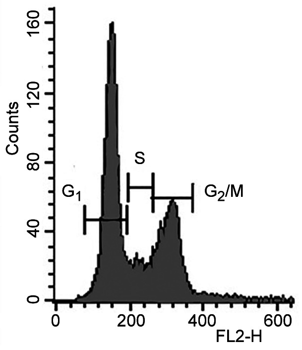
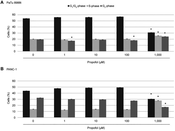
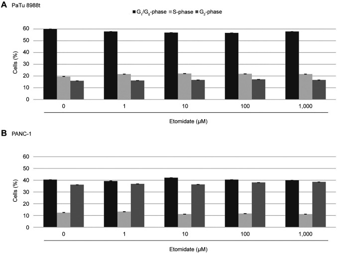
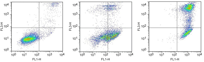
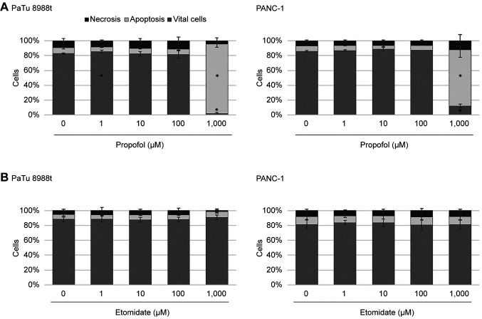
Materials and methods: To investigate the effect of propofol and etomidate on the proliferation, cell-cycle distribution, apoptosis, and necrosis of pancreatic tumour cells in vitro, PaTu 8988t and Panc-1 pancreatic cancer cells were treated with 0-1,000 μM propofol or etomidate for 24 h each. Cell proliferation was measured with enzyme-linked immunosorbent-bromodeoxyuridine assay. The apoptosis rate was analysed with annexin V staining and the cell-cycle distribution with flow cytometry.
Results: Propofol at 1,000 μM induced apoptosis and inhibited cell proliferation. The cell cycle showed an increased S-phase and reduced cells in the G1-phase. At 100 μM, propofol significantly inhibited proliferation of the pancreatic cancer cell line PaTu 8988t and reduced cells in the G2-phase in the cell cycle. Etomidate had no effects on cell-cycle distribution, proliferation, apoptosis, and necrosis at the concentrations used.
Conclusion: In this study, propofol was shown to have anticancer effects by induction of apoptosis and inhibition of cell proliferation, while etomidate did not affect pancreatic cancer cells. However, it is too early to make any recommendation for changes in clinical practice and further clinical studies are warranted to investigate the effect of anaesthetics on cancer progression.
Keywords: Propofol; apoptosis; cancer; cell-cycle distribution; etomidate; necrosis; pancreatic cancer; proliferation.
Copyright © 2022, International Institute of Anticancer Research (Dr. George J. Delinasios), All rights reserved.
Conflict of interest statement
The Authors declare that they have no competing interests.
Figures
References
-
- Gaylord HR, Simpson BT. The effect of certain anesthetics and loss of blood upon the growth of transplanted mouse cancer. J Can Res. 1916;1:379–380.
MeSH terms
Substances
LinkOut - more resources
Full Text Sources
Medical