. 2023 Mar;159(3):275-292.
doi: 10.1007/s00418-022-02156-3. Epub 2022 Oct 29.

In vivo ligamentogenesis in embroidered poly(lactic-co-ε-caprolactone) / polylactic acid scaffolds functionalized by fluorination and hexamethylene diisocyanate cross-linked collagen foams

Affiliations

In vivo ligamentogenesis in embroidered poly(lactic-co-ε-caprolactone) / polylactic acid scaffolds functionalized by fluorination and hexamethylene diisocyanate cross-linked collagen foams

Maria Kokozidou et al. Histochem Cell Biol. 2023 Mar.

Abstract

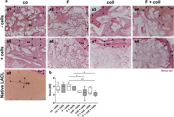
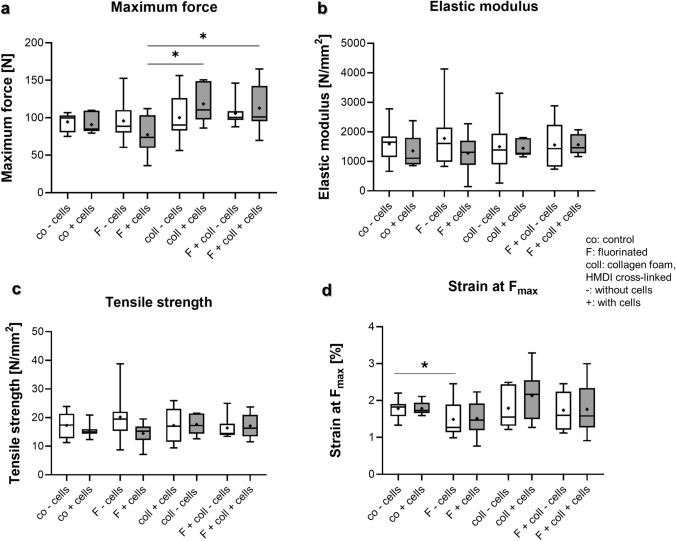
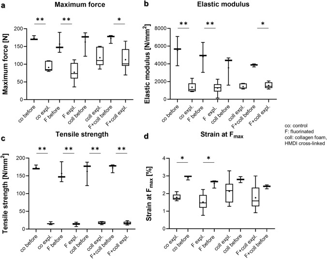
Although autografts represent the gold standard for anterior cruciate ligament (ACL) reconstruction, tissue-engineered ACLs provide a prospect to minimize donor site morbidity and limited graft availability. This study characterizes the ligamentogenesis in embroidered poly(L-lactide-co-ε-caprolactone) (P(LA-CL)) / polylactic acid (PLA) constructs using a dynamic nude mice xenograft model. (P(LA-CL))/PLA scaffolds remained either untreated (co) or were functionalized by gas fluorination (F), collagen foam cross-linked with hexamethylene diisocyanate (HMDI) (coll), or F combined with the foam (F + coll). Cell-free constructs or those seeded for 1 week with lapine ACL ligamentocytes were implanted into nude mice for 12 weeks. Following explantation, cell vitality and content, histo(patho)logy of scaffolds (including organs: liver, kidney, spleen), sulphated glycosaminoglycan (sGAG) contents and biomechanical properties were assessed.Scaffolds did not affect mice weight development and organs, indicating no organ toxicity. Moreover, scaffolds maintained their size and shape and reflected a high cell viability prior to and following implantation. Coll or F + coll scaffolds seeded with cells yielded superior macroscopic properties compared to the controls. Mild signs of inflammation (foreign-body giant cells and hyperemia) were limited to scaffolds without collagen. Microscopical score values and sGAG content did not differ significantly. Although remaining stable after explantation, elastic modulus, maximum force, tensile strength and strain at Fmax were significantly lower in explanted scaffolds compared to those before implantation, with no significant differences between scaffold subtypes, except for a higher maximum force in F + coll compared with F samples (in vivo). Scaffold functionalization with fluorinated collagen foam provides a promising approach for ACL tissue engineering. a Lapine anterior cruciate ligament (LACL): red arrow, posterior cruciate ligament: yellow arrow. Medial anterior meniscotibial ligament: black arrow. b Explant culture to isolate LACL fibroblasts. c Scaffold variants: co: controls; F: functionalization by gas-phase fluorination; coll: collagen foam cross-linked with hexamethylene diisocyanate (HMDI). c1-2 Embroidery pattern of the scaffolds. d Scaffolds were seeded with LACL fibroblasts using a dynamical culturing approach as depicted. e Scaffolds were implanted subnuchally into nude mice, fixed at the nuchal ligament and sacrospinal muscle tendons. f Two weeks after implantation. g Summary of analyses performed. Scale bars 1 cm (b, d), 0.5 cm (c). (sketches drawn by G.S.-T. using Krita 4.1.7 [Krita foundation, The Netherlands]).

Keywords: Anterior cruciate ligament; Dynamic nude mice xenograft model; Embroidered; Ligamentogenesis; Poly(L-lactide-co-ε-caprolactone) (P(LA-CL)); Polylactic acid (PLA) scaffolds; Tissue engineering.


Conflict of interest statement

The authors declare no competing interests.

All authors declared that they have no known competing financial interests or personal relationships that could have appeared to influence the work reported in this paper.

Figures

Fig. 1
Fig. 1
Body weight development of the animals, weight, and macroscopical appearance of the scaffold variants before implantation (after 1 week in vitro) and after explantation after 3 months in vivo. a1 Mice with a scaffold implanted into the subnuchal region. b1 Body weight of the mice in the week before scaffold implantation (red box plots) and in the week of explantation (black box plots). b2 Scaffold weight (wet weights, red box plots) pre-implantation and post explantation (black box plots). Macroscopical appearance of the lapine anterior cruciate ligament (LACL) (c1, red arrow) and the scaffold variants before implantation (c2–c5) and after explantation (c6–c13). d Results of macroscoring of the scaffolds. Co: controls; F: functionalization by gas fluorination; coll: collagen foam cross-linked with HMDI, -: implanted without cells, + : implanted with LACL-derived ligamentocytes, cultured for 1 week on the scaffold before scaffold implantation. p values: * < 0.05, ** < 0.01. Scale bars 1 cm
Fig. 2
Fig. 2
Cell viability and content in the scaffolds before implantation and after explantation after 3 months remaining in vivo. Cell viability in the scaffold variants before implantation after 1 week (a1–a4) and after explantation after 3 months in the nude mice model (a5–a12), scaffolds implanted without cells (a5–a8), and with lapine anterior cruciate ligament (LACL)-derived ligamentocytes, cultured for 1 week on the scaffold before scaffold implantation (a9–a12), control (a1, a5, a9), F (a2, a6, a10), collagen (a3, a7, a11) and F + collagen (a4, a8, a12). Living cells are stained green and dead cells are stained in red. b Measurement of DNA contents (pg DNA per cm3 scaffold) of the scaffold variants after implantation. Co: controls; F: functionalization by gas-phase fluorination; coll: collagen foam cross-linked with hexamethylene diisocyanate (HMDI), -: implanted without cells, + : implanted with LACL-derived ligamentocytes. Scale bar 100 µm
Fig. 3
Fig. 3
Scaffold histology after explantation after 3 months in vivo depicted by HE staining. Histology of the tissue is shown within the scaffold variants implanted without cells (-cells, a1–a4) and with lapine anterior cruciate ligament (LACL)-derived ligamentocytes (+ cells, a5–a8). Control (co, a1, a5), functionalization by gas-phase fluorination (F, a2, a6), collagen foam cross-linked with hexamethylene diisocyanate (HMDI) (coll, a3, a7) and F + coll (a4, a8). Native LACL (a9). Foreign-body giant cell and inflammatory cells visible in a co-cells scaffold variant (a10, FBGC). -cells: implanted without cells, + cells: implanted with LACL-derived ligamentocytes. BV blood vessel, PLA poly lactic acid, P(LA-CL) poly(lactic-co-ε-caprolactone). Double-headed black arrows capsule. In all images, the outer capsule surrounding the scaffold variants can be expected at the upper border. Scale bar 200 µm (a1–a9), 250 µm (a10)
Fig. 4
Fig. 4
Visualization of sulphated glycosaminoglycans in the different scaffold variants explanted after 3 months in vivo. AB stain to visualize sulphated glycosaminoglycans (sGAGs) of the tissue within the scaffold variants without cells (-cells, a1–a4) and with lapine anterior cruciate ligament (LACL)-derived ligamentocytes, cultured for 1 week on the scaffold in vitro before implantation into nude mice for 12 weeks (a5–a8), control (co, a1, a5), functionalization by gas-phase fluorination (F, a2, a6), collagen foam cross-linked with hexamethylene diisocyanate (HMDI) (coll, a3, a7) and F + coll (a4, a8). AB-stained native LACL (a9). b Results of AB scoring. c sGAG content measured by dimethyl methylene blue (DMMB) assay. -: implanted without cells, + : implanted with LACL-derived ligamentocytes. Kruskal–Wallis test was used for analyses followed by Dunn's post hoc test. p values: * < 0.05. Scale bars 100 µm (a1–a9)
Fig. 5
Fig. 5
Visualization of collagen in the different scaffold variants explanted after 3 months in vivo. SR stain to visualize collagen organization of the tissue within the scaffold variants without cells (a1–a4) and implanted with lapine anterior cruciate ligament (LACL)-derived ligamentocytes, cultured for 1 week on the scaffold in vitro before explanted after 12 weeks in vivo (a5–a8), co (a1, a5), F (a2, a6), coll (a3, a7) and F + coll (a4, a8). Native LACL (a9). Double-headed black arrows indicate an envelope rich in collagen surrounding the scaffold. b Results of Sirius red scoring. -: implanted without cells, + : implanted with LACL-derived ligamentocytes. Ad adipocytes, BV blood vessels, Epi epiligament, IFM interfascicular matrix. *prolene thread used for scaffold fixation. Kruskal–Wallis test was used for analyses followed by Dunn's post hoc test. p values: * < 0.05. Scale bars 400 µm (a1–a9)
Fig. 6
Fig. 6
Biomechanical analyses of scaffold variants explanted after 3 months in vivo. a Maximum force, b elastic modulus, c tensile strength, d strain at maximum force. Co controls, F functionalization by gas fluorination, coll collagen foam cross-linked with hexamethylene diisocyanate (HMDI), -: implanted without cells, + : implanted with lapine anterior cruciate ligament (LACL)-derived ligamentocytes, cultured for 1 week on the scaffold before scaffold implantation. Normality was assessed using a Shapiro–Wilk test and groups compared using Kruskal–Wallis test. p values: * < 0.05
Fig. 7
Fig. 7
Biomechanical analyses of scaffold variants explanted after 3 months in vivo compared to those before implantation (cultured 1 week in vitro). a Maximum force, b elastic modulus, c tensile strength, d strain at maximum force. Unseeded scaffolds and those seeded with cells were pooled. Co controls, F functionalization by gas-phase fluorination; coll collagen foam cross-linked with hexamethylene diisocyanate (HMDI). Normality was assessed using a Shapiro–Wilk test and groups compared using Kruskal–Wallis test. p values: * < 0.05, ** < 0.01, *** < 0.005

Similar articles

Cited by

References

    1. Archer DE, Mafi R, Mafi P, Khan WS. Preclinical studies on biomaterial scaffold use in knee ligament regeneration: a systematic review. Curr Stem Cell Res Ther. 2018;13:691–701. doi: 10.2174/1574888X13666180809093343. - DOI - PubMed
    1. Breier AC. Embroidery technology for hard-tissue scaffolds. Biomedical Textiles for Orthopaedic and Surgical Applications: Fundamentals, Applications and Tissue Engineering, Woodhead Publishing Series in Biomaterials; 2015. pp. 23–43.
    1. Cédric Laurent XL, De Isla N, Wang X, Rahouadj R. Defining a scaffold for ligament tissue engineering: What has been done, and what still needs to be done. J Cellular Immunotherapy. 2018;4:4–9. doi: 10.1016/j.jocit.2018.09.002. - DOI
    1. Chang CW, Lee JH, Chao PG. Chemical optimization for functional ligament tissue engineering. Tissue Eng Part A. 2020;26:102–110. doi: 10.1089/ten.tea.2019.0142. - DOI - PubMed
    1. Dai L, Hu X, Zhang X, Zhu J, Zhang J, Fu X, Duan X, Ao Y, Zhou C. Different tenogenic differentiation capacities of different mesenchymal stem cells in the presence of BMP-12. J Transl Med. 2015;13:200. doi: 10.1186/s12967-015-0560-7. - DOI - PMC - PubMed

LinkOut - more resources