. 2022 Oct 29;20(1):462.
doi: 10.1186/s12951-022-01674-5.

Nanoparticle-mediated selective Sfrp-1 silencing enhances bone density in osteoporotic mice

Affiliations

Nanoparticle-mediated selective Sfrp-1 silencing enhances bone density in osteoporotic mice

Patricia García-García et al. J Nanobiotechnology. .

Abstract

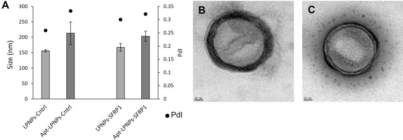
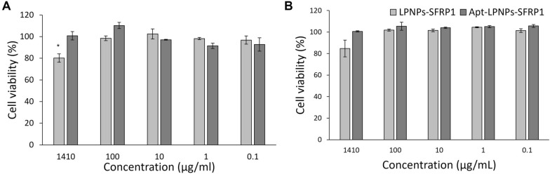
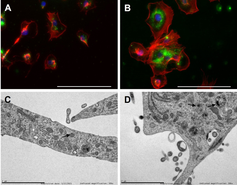
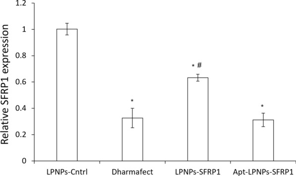
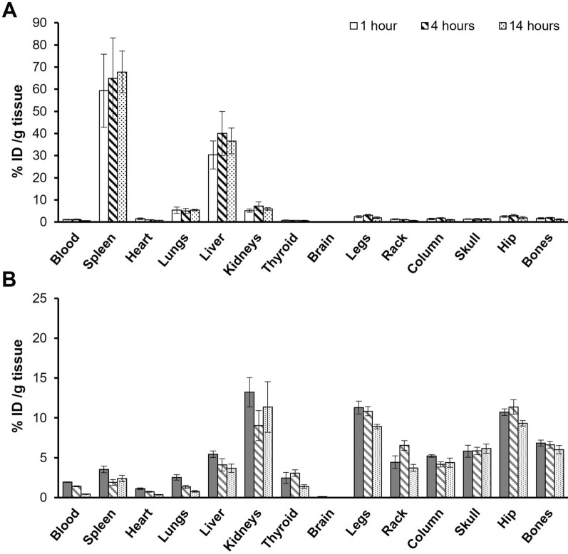
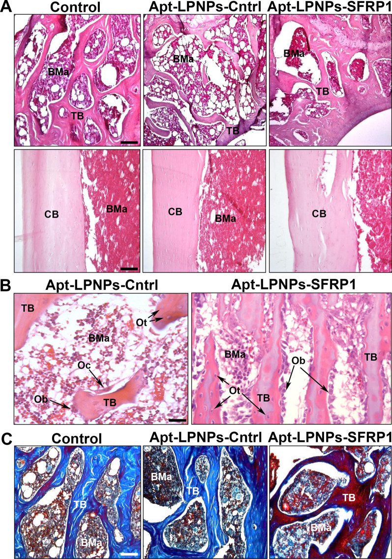
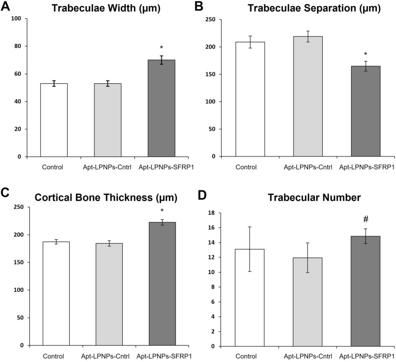
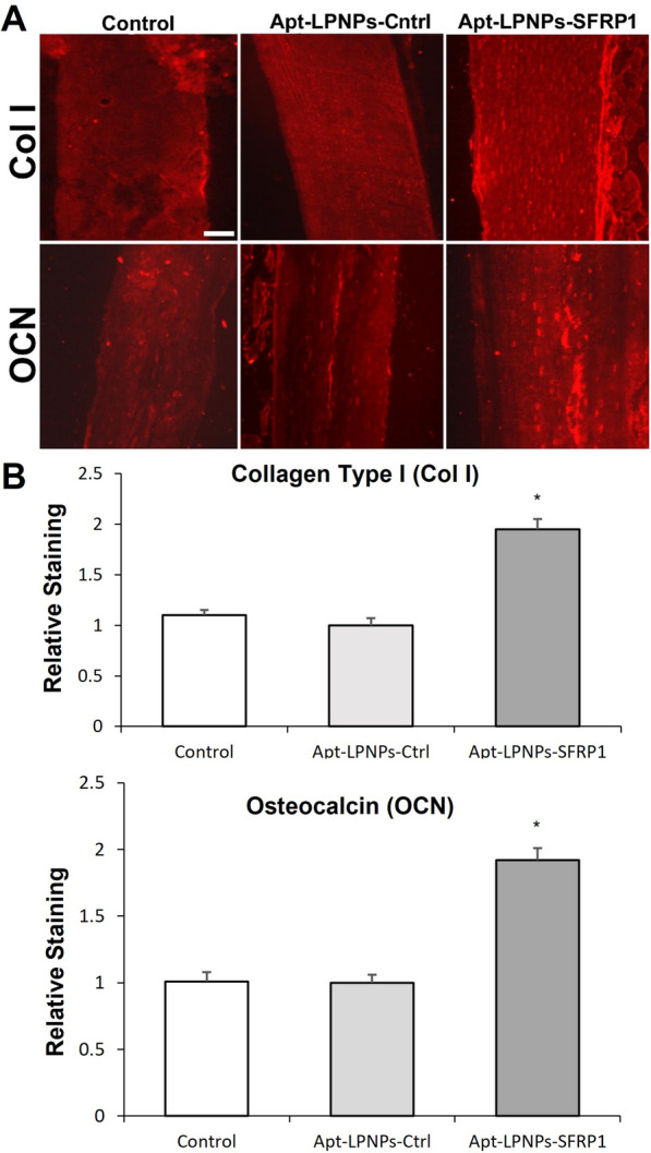
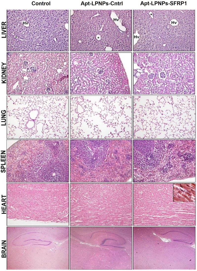
Osteoporosis (OP) is characterized by a loss in bone mass and mineral density. The stimulation of the canonical Wnt/β-catenin pathway has been reported to promote bone formation, this pathway is controlled by several regulators as secreted frizzled-related protein-1 (Sfrp-1), antagonist of the pathway. Thus, Sfrp-1 silencing therapies could be suitable for enhancing bone growth. However, the systemic stimulation of Wnt/β-catenin has been correlated with side effects. This work hypothesizes the administration of lipid-polymer NPs (LPNPs) functionalized with a MSC specific aptamer (Apt) and carrying a SFRP1 silencing GapmeR, could favor bone formation in OP with minimal undesired effects. Suitable SFRP1 GapmeR-loaded Apt-LPNPs (Apt-LPNPs-SFRP1) were administered in osteoporotic mice and their biodistribution, toxicity and bone induction capacity were evaluated. The aptamer functionalization of the NPs modified their biodistribution profile showing a four-fold increase in the bone accumulation and a ten-fold decrease in the hepatic accumulation compared to naked LPNPs. Moreover, the histological evaluation revealed evident changes in bone structure observing a more compact trabecular bone and a cortical bone thickness increase in the Apt-LPNPs-SFRP1 treated mice with no toxic effects. Therefore, these LPNPs showed suitable properties and biodistribution profiles leading to an enhancement on the bone density of osteoporotic mice.

Keywords: Bone regeneration; Gene therapy; Lipid-polymer hybrid nanoparticles; Osteoporosis.


Conflict of interest statement

Not applicable.

Figures

Fig. 1
Fig. 1
LPNPs properties in phosphate buffer at different pHs 5.4, 6.0, 7.0, 7.4 or in water at native conditions. Histogram indicates the Zeta potential (mV) values at different conditions and black points indicates nanoparticle size (nm) (n = 3)
Fig. 2
Fig. 2
A LPNPs-Cntrl and LPNPs-SFRP1 size (nm) and polydispersity index before and after aptamer conjugation obtained by DLS and Transmission electron micrographs of B LPNPs-SRFP1 and C Apt-LPNPs-SFRP1
Fig. 3
Fig. 3
Cell viability of A primary murine bone marrow mesenchymal stem cells (mMSC) and B murine macrophages (Raw 264.7) after 24 h of treatment with the LPNPs suspensions at variable concentrations (1.41 mg/mL-0.1 µg/mL). (*) denotes statistical significance to control (non-treated cells) p < 0.05 (n = 3)
Fig. 4
Fig. 4
Fluorescent microscopy and transmission electron microscopy images of A, C LPNPs and B, D Apt-LPNPs. Nanoparticles are labeled in green, cytoplasm is marked in red and cell nuclei are stained in blue. In electron micrographs LPNPs and Apt-LPNPs are indicated with arrows. Scale bars: Fluorescent microscopy: 20 µm. Electron micrographs: 2 µm
Fig. 5
Fig. 5
SFRP1 relative expression normalized by LPNPs-Cntrl after housekeeping normalization. (*) denotes statistical significance to LPNPs-Cntrl, (#) denotes statistical significance to the positive control Dharmafect (p < 0.05)
Fig. 6
Fig. 6
Organ accumulation of A Naked LPNPs and B Apt-LPNPs after i.v. administration using 99mTc radiolabeled NPs. The “bones” value is the average of all the radioactivity measurements of the different bone (n = 4)
Fig. 7
Fig. 7
Histology. Representative images showing the trabecular bone microarchitecture (A, upper line) and the cortical bone structure (A, bottom line) in the three experimental groups (SS, A-A-LPNPs control and A-A-LPNPs). B Images at high magnification, showing details of the osteosynthesis cellular activity in the A-A-LPNPs control and A-A-LPNPs groups. Observe the hypertrophic appearance of the osteoblasts (Ob) in the GapmeR-SFRP1 group, indicative of a higher synthesis activity, compared to the scares hypertrophic osteoblasts (Ob) in the A-A-LPNPs control. C Representative images with VOF staining technique, showing the mineralization degree in the three experimental groups. Observe the red-brown staining of the trabecular bone in the A-A-LPNPs group, indicative of a bone with a higher degree of mineralization compared to the control groups (SS and A-A-LPNPs control). BMa: bone marrow, CB: cortical bone, Ob: osteoblast, Oc: osteoclast; Ot: osteocytes, TB: trabecular bone. (A and B: H-Er; C: VOF). Scale bars: A upper line: 70 µm; bottom line: 60 µm. B 40 µm. C 50 µm
Fig. 8
Fig. 8
Histomorphometric analysis of femurs from the different experimental groups Control (0.9% saline solution) Apt-LPNPs-Cntrl and Apt-LPNPs-SFRP1.Thickness of the cortical bone (Ct.Wi) (A) and number (Tb.N) (B), width (Tb.Wi) (C) and separation (Tb.Sp) (D) of the trabeculae in trabecular bone. Histograms represent mean ± SE values. (*) denotes statistical significance between Control and Apt-LPNPs-Cntrl to Apt-LPNPs-SFRP1, (#) denotes statistical significance between Apt-LPNPs-Cntrl to Apt-LPNPs-SFRP1 (p < 0.005) (n = 6)
Fig. 9
Fig. 9
Immunohistochemical analysis of the femur samples from the different experimental groups: Control (0.9% saline solution), Apt-LPNPs-Cntrl and Apt-LPNPs-SFRP1. A Semipanoramic images of cortical bone showing the presence of Col I and OCN immunoreactivity. Scale bar: 50 µm. B Graphs showing the relative staining intensity of Col I and OCN in the three experimental groups. Histograms represent mean ± SEM values. (*) denotes statistical significance between Control and Apt-LPNPs-Cntrl to Apt-LPNPs-SFRP1 (p < 0.05) (n = 6)
Fig. 10
Fig. 10
Organs Toxicity. Representative semi-panoramic images in the three experimental groups, Control (0.9% saline solution), Apt-LPNPs-Cntrl and Apt-LPNPs-SFRP1, showing the histological structure of the liver, kidney, lung, spleen, heart and brain. All the organs analyzed showed normal tissue architecture with no apparent changes at the cellular level. Liver: Hv: hepatic vein, PV: portal vein, arrowheads: sinusoid capillaries. Kidney: CR: renal corpuscle, TCP: proximal convoluted tubule, TCD: distal convoluted tubule, arrowhead: renal capsule. Lung: A: artery, B: bronchiole, RB: respiratory bronchiole, AS: alveolar sacs, PA: pulmonary alveoli. Spleen: A: artery, RP: red pulp, WP: white pulp, arrowhead: splenic capsule. Heart: c: cardiomyocyte, E: epicardium, M: myocardium, arrowhead: intercalar disc. Brain: BF: basalforebrain, Hy: hippocampus, TC: telencephalic cortex, V: cerebral ventricles with choroid plexuses. Scale bars: liver, kidney, lung, spleen and heart: 80 µm. Heart insert: 15 µm. Brain: 150 µm

References

    1. Yadav VK, Balaji S, Suresh PS, Liu XS, Lu X, Li Z, Guo XE, Mann JJ, Balapure AK, Gershon MD, et al. Pharmacological inhibition of gut-derived serotonin synthesis is a potential bone anabolic treatment for osteoporosis. Nat Med. 2010;16:308–312. doi: 10.1038/nm.2098. - DOI - PMC - PubMed
    1. Hernlund E, Svedbom A, Ivergård M, Compston J, Cooper C, Stenmark J, McCloskey EV, Jönsson B, Kanis JA. Osteoporosis in the European Union: medical management, epidemiology and economic burden: A report prepared in collaboration with the International Osteoporosis Foundation (IOF) and the European Federation of Pharmaceutical Industry Associations (EFPIA) Arch Osteoporos. 2013 doi: 10.1007/s11657-013-0136-1. - DOI - PMC - PubMed
    1. Houschyar KS, Tapking C, Borrelli MR, Popp D, Duscher D, Maan ZN, Chelliah MP, Li J, Harati K, Wallner C, et al. Wnt pathway in bone repair and regeneration—what do we know so far. Front Cell Dev Biol. 2018;6:170. doi: 10.3389/fcell.2018.00170. - DOI - PMC - PubMed
    1. Bodine PV, Zhao W, Kharode YP, Bex FJ, Lambert AJ, Goad MB, Gaur T, Stein GS, Lian JB, Komm BS. The Wnt antagonist secreted frizzled-related protein-1 is a negative regulator of trabecular bone formation in adult mice. Mol Endocrinol. 2004;18:1222–1237. doi: 10.1210/me.2003-0498. - DOI - PubMed
    1. Bodine PV, Stauffer B, Ponce-de-Leon H, Bhat RA, Mangine A, Seestaller-Wehr LM, Moran RA, Billiard J, Fukayama S, Komm BS, et al. A small molecule inhibitor of the Wnt antagonist secreted frizzled-related protein-1 stimulates bone formation. Bone. 2009;44:1063–1068. doi: 10.1016/j.bone.2009.02.013. - DOI - PubMed