. 2022 Oct 27;14(20):8321-8345.
doi: 10.18632/aging.204320. Epub 2022 Oct 27.

Jian-Pi-Yi-Shen decoction inhibits mitochondria-dependent granulosa cell apoptosis in a rat model of POF

Affiliations

Jian-Pi-Yi-Shen decoction inhibits mitochondria-dependent granulosa cell apoptosis in a rat model of POF

Xiao-Lin Jiang et al. Aging (Albany NY). .

Erratum in

Abstract

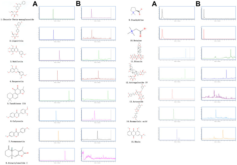
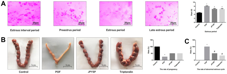
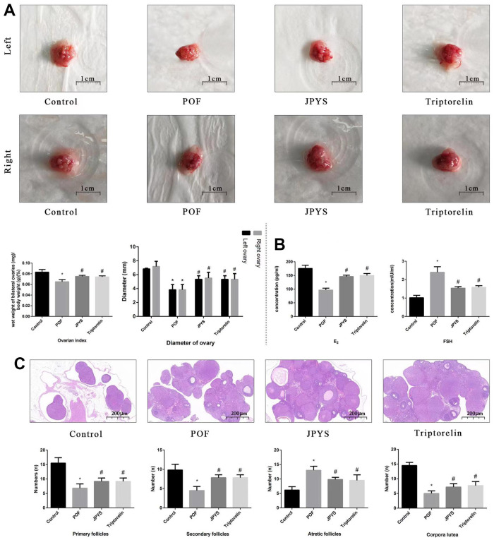
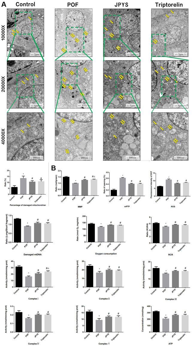
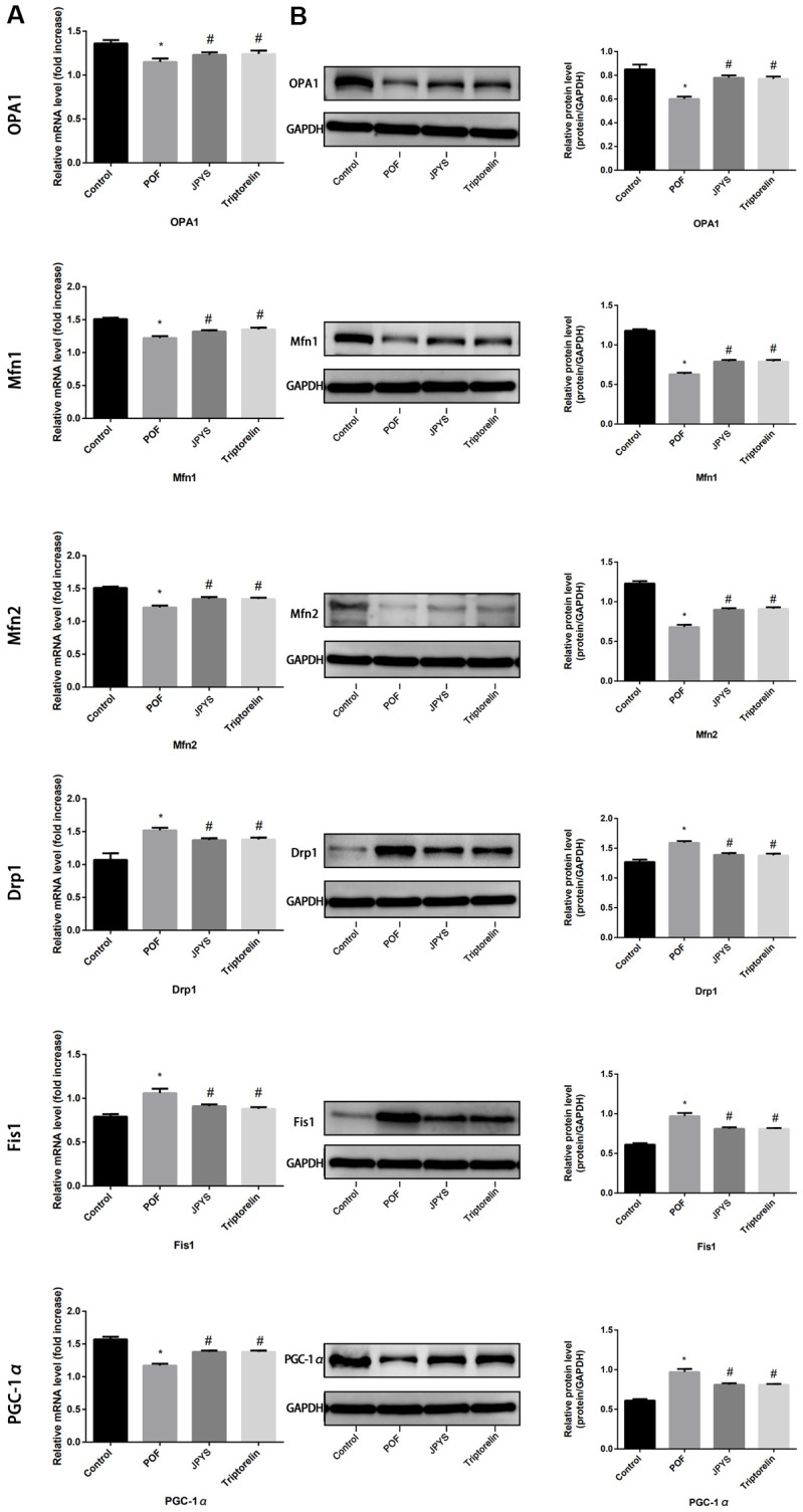
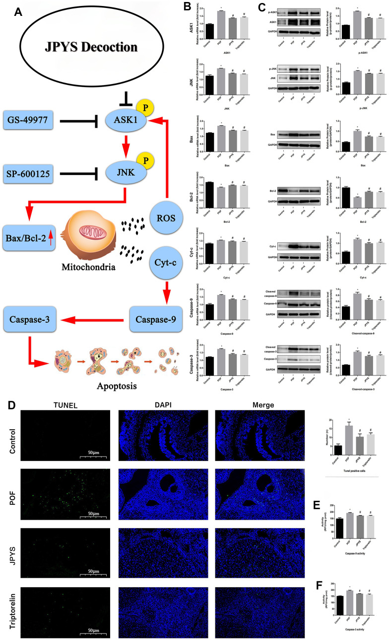
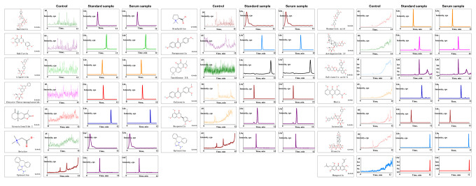
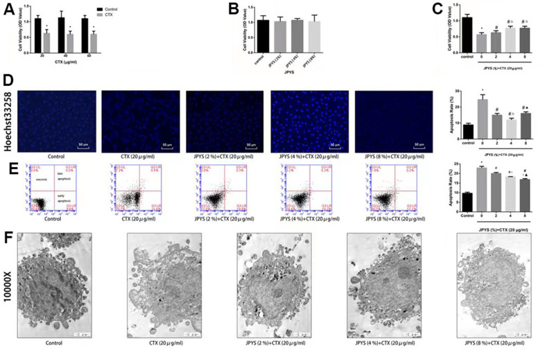
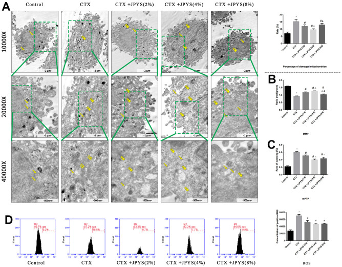
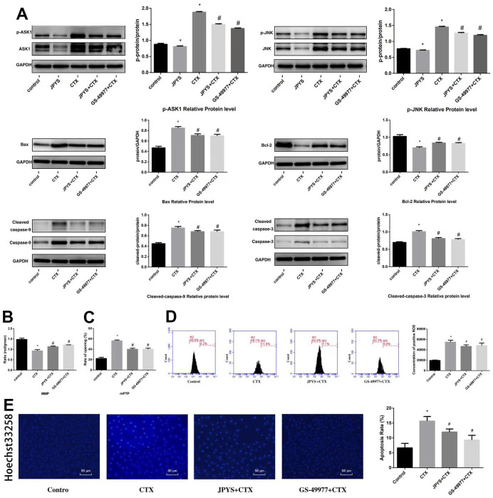
As a widely applied traditional Chinese medicine (TCM), Jian-Pi-Yi-Shen (JPYS) decoction maybe applied in curing premature ovarian failure (POF) besides chronic kidney disease (CKD). In vivo experiments, 40 female SD (8-week-old) rats were randomized into four groups, namely, control group (negative control), POF model group, JPYS treatment group, and triptorelin treatment group (positive control). JPYS group was treated with JPYS decoction (oral, 11 g/kg) for 60 days, and the triptorelin group was treated with triptorelin (injection, 1.5 mg/kg) for 10 days before the administration of cyclophosphamide (CTX) (50 mg/kg body weight) to establish POF model. We examined apoptosis, mitochondrial function, and target gene (ASK1/JNK pathway and mitochondrial fusion/fission) expression. In vitro experiments, the KGN human granulosa cell line was used. Cells were pretreated with CTX (20, 40, and 60 μg/mL) for 24 h, followed by JPYS-containing serum (2, 4, and 8 %) for 24 h. Thereafter, these cells were employed to assess apoptosis, mitochondrial function, and target gene levels of protein and mRNA. In vivo, JPYS alleviated injury and suppressed apoptosis in POF rats. In addition, JPYS improved ovarian function. JPYS inhibit apoptosis of granulosa cells through improving mitochondrial function by activating ASK1/JNK pathway. In vitro, JPYS inhibited KGN cell apoptosis through inhibited ASK1/JNK pathway and improved mitochondrial function. The effects of GS-49977 were similar to those of JPYS. During POF, mitochondrial dysfunction occurs in the ovary and leads to granulosa cell apoptosis. JPYS decoction improves mitochondrial function and alleviates apoptosis through ASK1/JNK pathway.

Keywords: Jian-Pi-Yi-Shen; apoptosis; granulosa cell; mitochondrial dysfunction; premature ovarian failure.


Conflict of interest statement

CONFLICTS OF INTEREST: The authors have no conflict of interest to disclose.

Figures

Figure 1
Figure 1
The extracted ion chromatograms of JPYS decoction. The extracted ion chromatograms of mixed standards (A); The extracted ion chromatograms of JPYS sample: 1.Calycosin-7-O-β-D-glucoside (CAS: 20633-67-4, MRM: 447.01/285.1, retention time: 4.343 min); 2. liquiritin (CAS: 551-15-5, MRM: 419.01/257.2, retention time: 4.463 min); 3. nobiletin CAS: 478-01-3, MRM: 403.01/373.2, retention time: 7.615 min); 4.hesperetin (CAS: 69097-99-0, MRM: 303.01/177.1, retention time: 6.591 min); 5. Tanshinone IIA (CAS: 568-72-9, MRM: 295.01/189.2, retention time: 10.986 min); 6.calycosin (CAS: 20575-57-9, MRM: 285.01/270.1, retention time: 5.767 min); 7. formononetin (CAS: 485-72-3, MRM: 269.01/197.1, retention time: 7.042 min); 8. atractylenolide I (CAS: 73069-13-3, MRM: 231.01/77.2, retention time: 10.185 min); 9. stachydrine (CAS: 471-87-4, MRM: 144.01/58.3, retention time: 1.000 min); 10. betaine (CAS: 478-01-3, MRM: 118.01/58.3, retention time: 1.092 min); 11. dioscin (CAS: 19057-60-4, MRM: 867.99/867.5, retention time: 8.656 min); 12. astragaloside IV (CAS: 96574-01-5, MRM: 783.99/783.5, retention time: 6.442 min); 13. acteoside (CAS: 61276-17-3, MRM: 622.99/161.1, retention time: 4.395 min); 14. rosmarinic acid (CAS: 20283-92-5, MRM: 358.99/161.1, retention time: 4.989 min); 15. rhein (CAS: 478-43-3, MRM: 282.99/239.1, retention time: 7.507 min) (B).
Figure 2
Figure 2
JPYS improved the abnormal estrous cycle and rate of pregnancy in premature ovarian failure (POF) rats. Rats were treated with JPYS (11.0 g/kg.d) and pre-treated with triptorelin (1.5 mg/kg) followed by intraperitoneally injected cyclophosphamide (50 mg/kg). (A) Estrous cycle of the rats (Estrous interval period: Vagina smear with white blood cells mainly; Preestrus period: With nuclear epithelial cells mainly; Estrous period: With Keratinized epithelial cells mainly; Late estrous period: See Keratinized epithelial cells and white blood cells) (n=6); (B) The rate of pregnancy (Female rats of each groups were sent to mate with male rats with ratio of 1:1 for 12 h. The mixture of sperm and vaginal smears seen on the next morning indicated the success of pregnancy, and this was considered as the 0.5 th day of the gestation. The pregnant rats were euthanized on the 13.5 th day of the gestation) (n=4); (C) The rate of abnormal estrous cycle (n=6). Data are shown as mean ± SD. *p < 0.05 versus control group, #p < 0.05 versus POF group, p < 0.05 versus JPYS group.
Figure 3
Figure 3
JPYS improved the ovarian function in premature ovarian failure (POF) rats. Rats were treated with JPYS (11.0 g/kg.d) and pre-treated with triptorelin (1.5 mg/kg) followed by intraperitoneally injected cyclophosphamide (50 mg/kg). (A) Diameter of bilateral ovaries and Ovarian index (Ovarian index= wet weight of bilateral ovaries (mg)/ body weight (g)×100 %, the size of the ovary in POF group were significantly reduced compared with control group), the scale bars represents a length of 1 cm on histology; (B) Ovarian function (follicle stimulating hormone (FSH) and oestradiol (E2)); (C) Histological assessment of the ovarian tissue using hematoxylin-eosin (HE) staining (All phases of follicles (primary follicles, secondary follicles, and atretic follicles) and corpora lutea were counted), the scale bars represents a length of 20 μm on histology. Data are shown as mean ± SD. *p < 0.05 versus control group, #p < 0.05 versus POF group, p < 0.05 versus JPYS group. (n=6).
Figure 4
Figure 4
JPYS improved mitochondrial function in premature ovarian failure (POF) rats. Rats were treated with JPYS (11.0 g/kg.d) and pre-treated with triptorelin (1.5 mg/kg) followed by intraperitoneally injected cyclophosphamide (50 mg/kg). (A) Electron microscope pictures (10,000×; 20,000×; 40 000×) of ovary in POF rats, the scale bars represents a length of 2 μm, 1 μm, and 500 nm on histology respectively. Abnormal mitochondrial (paired yellow arrow) morphology show that mitochondrial membrane rupture or swellings, normal mitochondrial (single yellow arrow) morphology type show that mitochondrial membrane smooth and inner carinulae distinct and percentage of damaged mitochondria; (B) The MMP (ratio of red/green), the opening of mPTP (%), the mitochondrial ROS, the mtDNA damage (ratio of long/short fragments), the mitochondrial RCR, mitochondrial oxygen consumption rate, the mitochondrial respiratory chain complex enzymes (I, II, III, IV, and V), and ATP were recorded above. Data are shown as mean ± SD. *p < 0.05 versus control group, #p < 0.05 versus POF group, p < 0.05 versus JPYS group. (n=6).
Figure 5
Figure 5
JPYS improved mitochondrial biogenesis and dynamics in premature ovarian failure (POF) rats. Rats were treated with JPYS (11.0 g/kg.d) and pre-treated with triptorelin (1.5 mg/kg) followed by intraperitoneally injected cyclophosphamide (50 mg/kg). We used real-time qPCR and western blot to detect mitochondrial biogenesis and dynamics. We chose OPA1, Mfn1, and Mfn2 to represent mitochondrial biogenesis function, and PGC-1α to represent the dynamic mitochondrial fusion, and Drp1 and Fis1 to represent mitochondrial fission. The expression of OPA1, Mfn1, Mfn2, PGC-1α, Drp1, and Fis1 in mRNA (A) and protein (B) levels. Data are shown as mean ± SD. *p < 0.05 versus control group, #p < 0.05 versus POF group, p < 0.05 versus JPYS group. (n=6).
Figure 6
Figure 6
JPYS improved mitochondrial function and inhibited apoptosis via inhibiting ASK1/JNK pathway. Rats were treated with JPYS (11.0 g/kg.d) and pre-treated with triptorelin (1.5 mg/kg) followed by intraperitoneally injected cyclophosphamide (50 mg/kg). Graphical abstract (A): Mitochondrial dysfunction of the ovarian GC occurs following POF and can lead to apoptosis. JPYS can relieve GC apoptosis by improving mitochondrial function via inhibiting the ASK1/JNK pathway in vivo and vitro. We used real-time qPCR and western blot to detect the target genes of ASK1/JNK pathway in mRNA (B) and protein (C) levels. TUNEL positive cells (D), the scale bars represents a length of 50 μm on histology. The activity of caspase-9 (E). The activity of caspase-3 (F). Data are shown as mean ± SD. *p < 0.05 versus control group, #p < 0.05 versus POF group, p < 0.05 versus JPYS group. (n=6).
Figure 7
Figure 7
Chemical component analysis of the JPYS-containing serum. Preparation of JPYS-Containing Serum. Female SD rats were intragastrically administrated with normal saline (control) or JPYS (11.0 g/kg/d) once daily for seven consecutive days. Then, 17 compounds (Rhein, salvianolic acid A, liquiritin, acteoside, calycosin-7-O-β-D-glucoside, rosmarinic acid, formononetin, calycosin, astragaloside IV, atractylenolide I, dioscin, tanshinone IIA, narirutin, nobiletin, hesperetin, stachydrine, betaine) were further quantified simultaneously by HPLC-QQQ-MS/MS.
Figure 8
Figure 8
JPYS improved decreased cell viability, increased cell apoptosis caused by cyclophosphamide (CTX) in vitro. JPYS-containing serum inhibited loss of cells viability induced by CTX. (A) Cells were treated with CTX (20, 40, and 60 μg/ml) for 24 h; (B) Cells were treated with JPYS-containing serum (2, 4, 8 %); (C) Cells were treated with CTX (20 μg/ml) for 24 h, then treated with JPYS-containing serum (2, 4, 8 %); (D) Hoechst 33258 staining was used to detect the apoptosis and counted the percentage of apoptotic cells; (E) Cell apoptosis was measured by fow cytometry and counted the percentage of apoptotic cells: provided 2-dimensional graphical representations of PI/annexin V-FITC plots. ‘Early apoptosis’ was defifined as cells positive for annexin V-FITC only. ‘Late apoptosis’ was defifined as cells positive for annexin V-FITC and PI. ‘Necrosis’ was defifined as cells positive for PI only; (F) Cell apoptosis was observed by electron microscope pictures (10,000×), the scale bars represents a length of 2 μm on cells respectively. Data are shown as mean ± SD. *p < 0.05 versus control group, #p < 0.05 versus CTX group, p < 0.05 versus CTX+ JPYS (2 %) group, p < 0.05 versus CTX+ JPYS (4 %) group. (n=3).
Figure 9
Figure 9
JPYS improved mitochondrial dysfunction caused by CTX in vitro. Cells were treated with CTX (20 μg/ml) for 24 h, then treated with JPYS-containing serum (2, 4, 8 %). (A) Electron microscope pictures (10,000×; 20,000×; 40,000×) of ovary in POF rats, the scale bars represents a length of 2 μm, 1 μm, and 500 nm on cells respectively. Abnormal mitochondrial (paired yellow arrow) morphology show that mitochondrial membrane rupture or swellings, normal mitochondrial (single yellow arrow) morphology type show that mitochondrial membrane smooth and inner carinulae distinct and percentage of damaged mitochondria; (B) The MMP (ratio of red/green); (C) The opening of the mPTP (%); (D) The ROS levels. Data are shown as mean ± SD. *p < 0.05 versus control group, #p < 0.05 versus CTX group, p < 0.05 versus CTX+ JPYS (2 %) group, p < 0.05 versus CTX+ JPYS (4 %) group. (n=3).
Figure 10
Figure 10
JPYS inhibited ASK1/JNK-induced apoptosis and mitochondrial dysfunction in vitro. To investigate whether the anti-apoptotic efect of JPYS-containing serum (4 %) was associated with the ASK1/JNK pathway. GS-49977 (5 μM) was the inhibitor of ASK1. (A) We used western blot to detect the target genes of the ASK1/JNK pathway in protein level; (B) The MMP (ratio of red/green); (C) The opening of mPTP (%); (D) The ROS levels; (E) Hoechst 33258 staining was used to detect the apoptosis and counted the percentage of apoptotic cells. Data are shown as mean ± SD. *p < 0.05 versus control group, #p < 0.05 versus CTX group. (n=3).

Similar articles

Cited by

References

    1. Kokcu A. Premature ovarian failure from current perspective. Gynecol Endocrinol. 2010; 26:555–62. 10.3109/09513590.2010.488773 - DOI - PubMed
    1. Grossmann B, Saur S, Rall K, Pecher AC, Hübner S, Henes J, Henes M. Prevalence of autoimmune disease in women with premature ovarian failure. Eur J Contracept Reprod Health Care. 2020; 25:72–5. 10.1080/13625187.2019.1702638 - DOI - PubMed
    1. Yuan S, Akey CW. Apoptosome structure, assembly, and procaspase activation. Structure. 2013; 21:501–15. 10.1016/j.str.2013.02.024 - DOI - PMC - PubMed
    1. Matsuda F, Inoue N, Manabe N, Ohkura S. Follicular growth and atresia in mammalian ovaries: regulation by survival and death of granulosa cells. J Reprod Dev. 2012; 58:44–50. 10.1262/jrd.2011-012 - DOI - PubMed
    1. Siristatidis C, Dafopoulos K, Salamalekis G, Galazios G, Christoforidis N, Moustakarias T, Koutlaki N, Bouschanetzis C, Loutradis D, Drakakis P. Administration of low-molecular-weight heparin in patients with two or more unsuccessful IVF/ICSI cycles: a multicenter cohort study. Gynecol Endocrinol. 2018; 34:747–51. 10.1080/09513590.2018.1442426 - DOI - PubMed

Publication types