Review
. 2023 Jan;51(1):111-122.
doi: 10.1124/dmd.122.001109. Epub 2022 Oct 30.

Four Decades of Cytochrome P450 2B Research: From Protein Adducts to Protein Structures and Beyond

Affiliations
Review

Four Decades of Cytochrome P450 2B Research: From Protein Adducts to Protein Structures and Beyond

Arthur G Roberts et al. Drug Metab Dispos. 2023 Jan.

Abstract

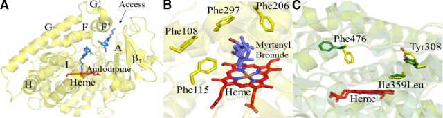
This article features selected findings from the senior author and colleagues dating back to 1978 and covering approximately three-fourths of the 60 years since the discovery of cytochrome P450. Considering the vast number of P450 enzymes in this amazing superfamily and their importance for so many fields of science and medicine, including drug design and development, drug therapy, environmental health, and biotechnology, a comprehensive review of even a single topic is daunting. To make a meaningful contribution to the 50th anniversary of Drug Metabolism and Disposition, we trace the development of the research in a single P450 laboratory through the eyes of seven individuals with different backgrounds, perspectives, and subsequent career trajectories. All co-authors are united in their fascination for the structural basis of mammalian P450 substrate and inhibitor selectivity and using such information to improve drug design and therapy. An underlying theme is how technological advances enable scientific discoveries that were impossible and even inconceivable to prior generations. The work performed spans the continuum from: 1) purification of P450 enzymes from animal tissues to purification of expressed human P450 enzymes and their site-directed mutants from bacteria; 2) inhibition, metabolism, and spectral studies to isothermal titration calorimetry, deuterium exchange mass spectrometry, and NMR; 3) homology models based on bacterial P450 X-ray crystal structures to rabbit and human P450 structures in complex with a wide variety of ligands. Our hope is that humanizing the scientific endeavor will encourage new generations of scientists to make fundamental new discoveries in the P450 field. SIGNIFICANCE STATEMENT: The manuscript summarizes four decades of work from Dr. James Halpert's laboratory, whose investigations have shaped the cytochrome P450 field, and provides insightful perspectives of the co-authors. This work will also inspire future drug metabolism scientists to make critical new discoveries in the cytochrome P450 field.


Figures

Fig. 1.
Fig. 1.
Metabolic scheme for bioactivation of chloramphenicol by CYP2B1 and release of the adduct N-ε-chloramphenicol oxamyl lysine upon proteolytic digestion of the modified enzyme.
Fig. 2.
Fig. 2.
Irreversible Type I spectral shift produced by the binding of 17α-ethynyl progesterone metabolite(s) to purified bovine adrenal CYP21A2 (Stevens et al., 1991). The figure was adapted with permission from Stevens JC, Jaw JY, Peng CT, and Halpert J, Mechanism-based inactivation of bovine adrenal cytochromes P450 C-21 and P450 17 alpha by 17 beta-substituted steroids. Biochemistry 30:3649–3658. Copyright 1991 American Chemical Society.
Fig. 3.
Fig. 3.
Docking of ligands into the active site of the CYP2B1 model. (A) Progesterone docked into the active site of the CYP2B1 L209A mutant in a binding orientation leading to 21-OH progesterone. Key amino acid residues are shown in purple; Ala-209 is in green. Leu-209 (purple) is present in the wild type (WT) enzyme, which leads to van der Waals overlaps with progesterone. (B) BBT (N-benzyl-1-aminobenzotriazole) docked into the active site of CYP 2B1 in an orientation allowing for oxidation at the 1-amino nitrogen, leading to enzyme inactivation. When Gly-478 is replaced with Ala, van der Waals overlaps appear between the inhibitor and the Ala side chain. Key residues are in purple, and Ala-478 is in green. Such overlaps likely account for the resistance of G478A to inactivation by the chloramphenicol analog N-(2-p-nitrophenethyl)chlorofluoroacetamide (He et al., 1992).
Fig. 4.
Fig. 4.
Comparison of cytochrome P450 2B4 structures in the open (green) and closed, ligand-bound (blue) states, highlighting highly mobile elements of the B’C loop (orange) and F’-G helices (purple).
Fig. 5.
Fig. 5.
Schematic representation of the directed evolution approach to engineer mammalian CYP2B1 enzyme.
Fig. 6.
Fig. 6.
My four research fields. During my research adventure, I investigated (A) Rieske (R) iron-sulfur domain movements with the cytochrome (cyt.) b6f complex, (B) drug–drug interactions with cytochrome P450 3A4 (CYP3A4), (C) transport with P-glycoprotein (P-gp), and (D) targeted plasmid delivery with bioengineered liposomes. PDB IDs: CYP3A4: 1TQN; cyt. b6f complex: 4PV1; P-gp: 5KPI; PC: 2BZC. Additional abbreviations: Ab, antibody; P.C., plastocyanin; PEG, polyethylene glycol; PS I, photosystem I. The figure was created with biorender.com.
Fig. 7.
Fig. 7.
Structures of human CYP2B6 and 2C9. (A) Dual ligand complex of CYP2B6 (Y226H/K262R) with amlodipine (violet sticks) bound that helped elucidate the substrate access channel from the active site near heme to the solvent region. (B) Structure of CYP2B6 (Y226H/K262R) bound with myrtenyl bromide (violet sticks) with bromine exhibiting halogen-π interaction with the aromatic phenylalanine side chains. (C) Structural overlay of CYP2C9 WT (green) and CYP2C9*3 (yellow) complexed with losartan. The *3 that represents isoleucine to leucine change at 359 affects neighboring tyrosine at 308 that impacts the residues in the active site including phenylalanine 476. The Phe576 side chain in *3 protrudes into the active site affecting the orientation of losartan compared with the WT complex. Losartan molecules are not shown for clarity purposes. Heme is shown in red stick representation.

References

    1. Bart AG, Scott EE (2017) Structural and functional effects of cytochrome b5 interactions with human cytochrome P450 enzymes. J Biol Chem 292:20818–20833. - PMC - PubMed
    1. Black SM, Harikrishna JA, Szklarz GD, Miller WL (1994) The mitochondrial environment is required for activity of the cholesterol side-chain cleavage enzyme, cytochrome P450scc. Proc Natl Acad Sci USA 91:7247–7251. - PMC - PubMed
    1. Brixius-Anderko S, Scott EE (2021) Aldosterone Synthase Structure With Cushing Disease Drug LCI699 Highlights Avenues for Selective CYP11B Drug Design. Hypertension 78:751–759. - PubMed
    1. Brixius-Anderko S, Scott EE (2019) Structure of human cortisol-producing cytochrome P450 11B1 bound to the breast cancer drug fadrozole provides insights for drug design. J Biol Chem 294:453–460. - PMC - PubMed
    1. Ciaccio PJ, Halpert JR (1989) Characterization of a phenobarbital-inducible dog liver cytochrome P450 structurally related to rat and human enzymes of the P450IIIA (steroid-inducible) gene subfamily. Arch Biochem Biophys 271:284–299. - PubMed

Publication types

Substances

LinkOut - more resources