. 2022 Oct 31;20(1):220.
doi: 10.1186/s12951-022-01400-1.

Bone-targeting delivery of platelet lysate exosomes ameliorates glucocorticoid-induced osteoporosis by enhancing bone-vessel coupling

Affiliations

Bone-targeting delivery of platelet lysate exosomes ameliorates glucocorticoid-induced osteoporosis by enhancing bone-vessel coupling

Gang Zheng et al. J Nanobiotechnology. .

Abstract

Background: Glucocorticoids (GCs) overuse is associated with decreased bone mass and osseous vasculature destruction, leading to severe osteoporosis. Platelet lysates (PL) as a pool of growth factors (GFs) were widely used in local bone repair by its potent pro-regeneration and pro-angiogenesis. However, it is still seldom applied for treating systemic osteopathia due to the lack of a suitable delivery strategy. The non-targeted distribution of GFs might cause tumorigenesis in other organs.

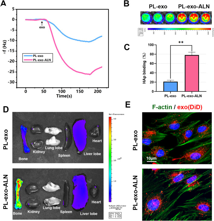
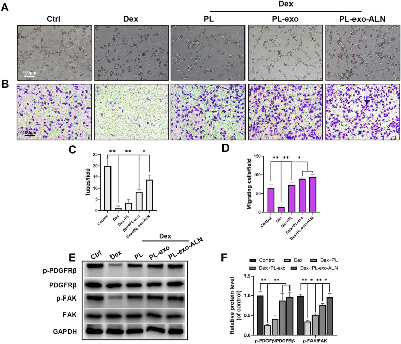
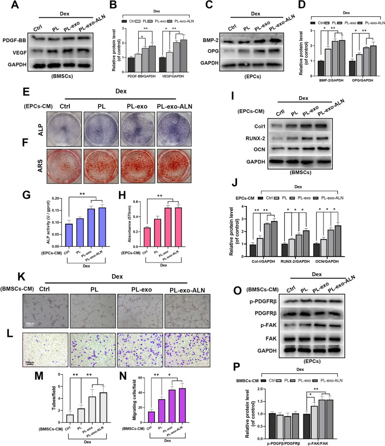
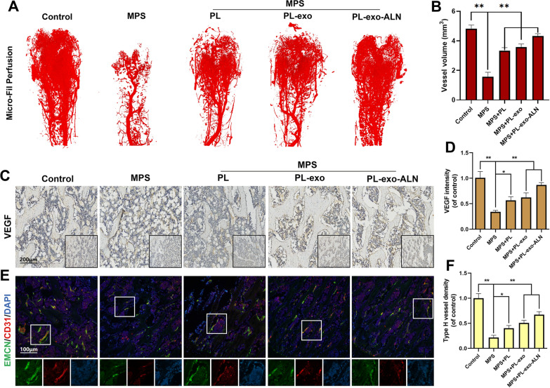
Results: In this study, PL-derived exosomes (PL-exo) were isolated to enrich the platelet-derived GFs, followed by conjugating with alendronate (ALN) grafted PEGylated phospholipid (DSPE-PEG-ALN) to establish a bone-targeting PL-exo (PL-exo-ALN). The in vitro hydroxyapatite binding affinity and in vivo bone targeting aggregation of PL-exo were significantly enhanced after ALN modification. Besides directly modulating the osteogenic and angiogenic differentiation of bone marrow mesenchymal stem cells (BMSCs) and endothelial progenitor cells (EPCs), respectively, PL-exo-ALN also facilitate their coupling under GCs' stimulation. Additionally, intravenous injection of PL-exo-ALN could successfully rescue GCs induced osteoporosis (GIOP) in vivo.

Conclusions: PL-exo-ALN may be utilized as a novel nanoplatform for precise infusion of GFs to bone sites and exerts promising therapeutic potential for GIOP.

Keywords: Bone-targeting; Exosome; Glucocorticoid; Osteoporosis; Platelet lysate.


Conflict of interest statement

The authors declare no conflict of interest.

Figures

Fig. 1
Fig. 1
Characteristics of the isolated PL-Exos. A Schematic illustration of PL-exo-ALN preparation and its bone targeting mechanism. B Representative TEM image of PL-exo and PL-exo-ALN (scale bar: 100 nm). C Representative DLS results showing the size distribution of PL-exo and PL-exo-ALN. D Confirmation of the presence of exosomal marker proteins (CD9 and TSG101), the decrease of platelet marker (CD41), the enrich of representative GFs (PDGF-BB, TGF-β, bFGF and VEGF) and the absence of the non-exosomal protein (calnexin) by Western blot analysis
Fig. 2
Fig. 2
Bone-targeting ability of PL-exo-ALN. A Frequency shifts of exosomes deposition on the HAp coated sensor were detected by QCM-D. B Fluorescent Image of DiD-labelled exosomes binding to HAp were detected by IVIS. C Quantitative analysis of DiD-labelled exosomes binding to HAp by a fluorescence microplate reader. D Fluorescent Image of the in vivo distribution of DiD-labelled exosomes by IVIS. E Internalization of exosomes into BMSCs (scale bar: 10 μm). All results are presented as the means ± SDs, *P < 0.05, **P < 0.01
Fig. 3
Fig. 3
Effects of PL, PL-exo and PL-exo-ALN on Dex-induced osteogenesis inhibition in BMSCs. AB Early osteogenic differentiation was determined by ALP staining and ALP activity assays after 5 days of induction. CD Late osteogenic differentiation was determined by Alizarin Red staining and the calcium deposition was quantified by measuring the optical density, after 14 days of induction. EF The expression levels of specific proteins in BMSCs treated as indicated for 3 days. G Immunofluorescence staining of Col-I (green), F-actin(red) and nucleus (blue) after 3 days induction (scale bar: 25 μm). All results are presented as the means ± SDs, *P < 0.05, **P < 0.01
Fig. 4
Fig. 4
Effects of PL, PL-exo and PL-exo-ALN on Dex-induced angiogenesis inhibition in EPCs. A In vitro tube formation assay of EPCs treated as indicated. (scale bar: 150 μm). B The migration evaluation of EPCs treated as indicated by transwell assay (scale bar: 100 μm). C Quantification of tube formation. D Quantification of migrated cells. EF The phosphorylation levels of PDGFRβ and FAK in EPCs treated as indicated for 24 h. All results are presented as the means ± SDs, *P < 0.05, **P < 0.01
Fig. 5
Fig. 5
The cross-talk between BMSCs and EPCs under the Dex-stimulated condition after treatment. AB The levels of PDGF-BB and VEGF in Dex-stimulated BMSCs treated as indicated for 24 h. CD The levels of BMP-2 and OPG in Dex-stimulated EPCs treated as indicated for 3 days. E, G Early osteogenic differentiation of BMSCs was determined by ALP staining and ALP activity assays after 5 days of induction. F, H Late osteogenic differentiation of BMSCs was determined by Alizarin Red staining and the calcium deposition was quantified by measuring the optical density, after 14 days of induction. IJ The expression levels of specific proteins in BMSCs treated as indicated for 3 days. K In vitro tube formation assay of EPCs treated as indicated (scale bar: 150 μm). L The migration evaluation of EPCs treated as indicated by transwell assay (scale bar: 100 μm). M Quantification of tube formation. N Quantification of migrated cells. OP The phosphorylation levels of PDGFRβ and FAK in EPCs treated as indicated for 24 h. All results are presented as the means ± SDs, *P < 0.05, **P < 0.01
Fig. 6
Fig. 6
Effects of PL, PL-exo and PL-exo-ALN on MPS-induced bone mass loss in vivo. A 2D and 3D reconstructive μ-CT images in the distal femurs of five groups. B Quantification of the bone mineral density (BMD), trabecular bone volume fraction (BV/TV), trabecular number (Tb.N), trabecular thickness (Tb.Th) and trabecular separation (Tb.Sp) by CTAn of five groups. C Images of dynamic bone formation with different treatments were monitored the fluorochrome labeling of five groups (scale bar: 300 μm). D Bone minerals of different groups were examined by Von Kossa staining of five groups. All results are presented as the means ± SDs, *P < 0.05, **P < 0.01
Fig. 7
Fig. 7
Effects of PL, PL-exo and PL-exo-ALN on MPS-induced fatty infiltration, down-regulation of osteogenic markers. A H&E staining of the distal femurs (scale bar: 300 μm). B Quantification of fat tissue on bone marrow. CF Images and semi-quantifications for IHC staining of Col-I and OCN (scale bar: 200 μm). G TRAcP staining of the distal femurs (scale bar: 200 μm). H Quantitation of Oc.S/BS. All results are presented as the means ± SDs, *P < 0.05, **P < 0.01
Fig. 8
Fig. 8
Effects of PL, PL-exo and PL-exo-ALN on MPS-induced vasculature destruction and decrease of angiogenic marker and H-type vessels. AB Vessel perfusion and related quantification of five groups as indicated. CD Images and semi-quantifications for IHC staining of VEGF. EF Immunofluorescence staining of EMCN (green), CD31 (red) and nucleus (blue) (scale bar: 100 μm). All results are presented as the means ± SDs, *P < 0.05, **P < 0.01

References

    1. Rentero ML, Amigo E, Chozas N, Prada MF, Silva-Fernández L, Hernandez MAA, Barrera JMR, del Pino-Montes J. Prevalence of fractures in women with rheumatoid arthritis and/or systemic lupus erythematosus on chronic glucocorticoid therapy. BMC Musculoskelet Disord. 2015;16:1–10. doi: 10.1186/s12891-015-0733-9. - DOI - PMC - PubMed
    1. Frenkel B, White W, Tuckermann J. Glucocorticoid-induced osteoporosis. Adv Exp Med Biol. 2015;872:179–215. doi: 10.1007/978-1-4939-2895-8_8. - DOI - PMC - PubMed
    1. Hansen KE, Kleker B, Safdar N, Bartels CM. A systematic review and meta-analysis of glucocorticoid-induced osteoporosis in children.Seminars in arthritis and rheumatism. Elsevier:Amsterdam; 2014.p. 47–54. - PMC - PubMed
    1. Iacopo C, Alberto F, Daniela M, Cristina EV, Luigi G. Updates in epidemiology, pathophysiology and management strategies of glucocorticoid-induced osteoporosis. Expert Rev Endocrinol Metab. 2020;15:283–298. doi: 10.1080/17446651.2020.1772051. - DOI - PubMed
    1. Lane NE. Glucocorticoid-induced osteoporosis: new insights into the pathophysiology and treatments. Curr Osteoporos Rep. 2019;17:1–7. doi: 10.1007/s11914-019-00498-x. - DOI - PMC - PubMed