. 2022 Oct 31;7(1):130.
doi: 10.1038/s41541-022-00556-z.

B cells oppose Mycoplasma pneumoniae vaccine enhanced disease and limit bacterial colonization of the lungs

Affiliations

B cells oppose Mycoplasma pneumoniae vaccine enhanced disease and limit bacterial colonization of the lungs

Tyler D Gavitt et al. NPJ Vaccines. .

Abstract

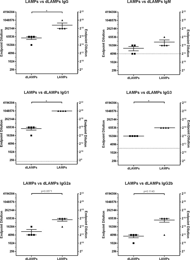
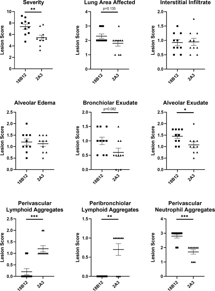
Development of an effective vaccine for Mycoplasma pneumoniae has been hindered by reports of Vaccine Enhanced Disease (VED) in test subjects vaccinated and challenged in studies conducted in the 1960s. The exact mechanism of disease exacerbation has yet to be fully described, but host immune responses to Lipid-Associated Membrane Proteins (LAMPs) lipoprotein lipid moieties have been implicated. LAMPs-induced exacerbation appears to involve helper T cell recall responses, due in part to their influence on neutrophil recruitment and subsequent inflammatory responses in the lung. Herein, we characterized the functions of host B cell responses to M. pneumoniae LAMPs and delipidated-LAMPs (dLAMPs) by conducting passive transfer and B cell depletion studies to assess their contribution to disease exacerbation or protection using a BALB/c mouse model. We found that antibody responses to M. pneumoniae LAMPs and dLAMPs differ in magnitude, but not in isotype or subclass. Passive transfer, dLAMP denaturation, and monoclonal antibody studies indicate that antibodies do not cause VED, but do appear to contribute to control of bacterial loads in the lungs. Depletion of B cells prior to LAMPs-vaccination results in significantly enhanced pathology in comparison to B cell competent controls, suggesting a possible regulatory role of B cells distinct from antibody secretion. Taken together, our findings suggest that B cell antibody responses to M. pneumoniae contribute to, but are insufficient for protection against challenge on their own, and that other functional properties of B cells are necessary to limit exacerbation of disease in LAMPs-vaccinated mice after infection.


Conflict of interest statement

The authors declare no competing interests.

Figures

Fig. 1
Fig. 1. Serum antibody responses to Lipid-associated Membrane Proteins (LAMPs) or delipidated Lipid-associated Membrane Proteins (dLAMPs) in vaccinated mice.
Endpoint titers are displayed as the reciprocal of the final dilution at which signal was at least three times greater than the assay background. Data were analyzed by Mann–Whitney Test. Data were considered significant for p < 0.05. Data without significance marking is to be considered not significantly different. Dotted line indicates the lower limit of detection of the assay.
Fig. 2
Fig. 2. Passive transfer of hyper-immune sera to naïve mice.
A Histopathologic scoring of lungs from mice intranasally inoculated with Lipid-associated Membrane Protein-induced (LAMP), delipidated Lipid-associated Membrane Protein-induced (dLAMP) or naïve-mouse (sham) serum at high (Hi) or low (Lo) concentration and challenged with M. pneumoniae. Data were compared by Kruskal–Wallis test with Dunn’s post-hoc test used for multiple comparisons. B Bacterial recoveries from the lungs of mice intranasally inoculated with vaccine-induced or naïve-mouse (Sham) serum at high or low concentration and challenged with M. pneumoniae. Data are displayed as median with interquartile range, and were compared by Kruskal–Wallis test with Dunn’s post-hoc test used for multiple comparisons. C Histopathologic scoring of lungs from mice intraperitoneally inoculated with vaccine-induced or naïve-mouse (sham) serum and challenged with M. pneumoniae. Data were compared by Kruskal–Wallis test with Dunn’s post-hoc test used for multiple comparisons. D Bacterial recoveries from the lungs of mice intraperitoneally inoculated with vaccine-induced or naïve-mouse (Sham) serum and challenged with M. pneumoniae. Data are displayed as median with interquartile range, and were compared by Kruskal–Wallis test with Dunn’s post-hoc test used for multiple comparisons.
Fig. 3
Fig. 3. Passive transfer of anti-P1 monoclonal antibody to naïve mice.
A Histopathologic scoring of lungs from mice intranasally inoculated with a-P1 monoclonal antibody and challenged with M. pneumoniae. Data points were compared by Mann–Whitney Test. B Bacterial recoveries from the lungs of mice intranasally inoculated with a-P1 monoclonal antibody one day before challenge (D-1) or one day after challenge (D + 1) with M. pneumoniae. Data are displayed as median with interquartile range. Data points were compared by Mann–Whitney Test. C Histopathologic scoring of lungs from mice intraperitoneally inoculated with a-P1 monoclonal antibody and challenged with M. pneumoniae. Data were compared by Kruskal–Wallis test with Dunn’s post-hoc test used for multiple comparisons. D Bacterial recoveries from the lungs of mice intraperitoneally inoculated with a-P1 monoclonal antibody and challenged with M. pneumoniae. Data are median with interquartile range. Data compared by Kruskal–Wallis test with Dunn’s post-hoc test used for multiple comparisons. Differences for all comparisons were considered significant for p < 0.05 (*p < 0.05, **p < 0.01, ***p < 0.001).
Fig. 4
Fig. 4. Vaccination with native or denatured dLAMPs.
A Histopathology scoring of lungs from mice intraperitoneally vaccinated with native conformation (NdL) or denatured (DdL) dLAMPs and then intranasally challenged with M. pneumoniae. Data were compared by Mann–Whitney Test and are displayed as median with interquartile range. B Bacterial recovery from lungs from mice intraperitoneally inoculated with NdL or DdL and intranasally challenged with M. pneumoniae. Data points were compared by Mann–Whitney Test, and differences were considered significant for p < 0.05 (***p < 0.001).
Fig. 5
Fig. 5. Pathologic effects of B cell depletion on LAMP vaccination and infection.
Histologic outcomes of mice vaccinated with M. pneumoniae LAMPs and pre-treated with the B cell depleting antibody 18B12.1 or the isotype control antibody 2A3. Severity score is the overall sum of interstitial infiltrate, alveolar exudate, alveolar edema, bronchiolar exudate, and perivascular neutrophil aggregate scores for individual animals. Data were analyzed using the Mann–Whitney test, displayed as mean plus and minus standard error of the mean, and differences were considered significant for p < 0.05 (*p < 0.05, **p < 0.01, ***p < 0.001).
Fig. 6
Fig. 6. In situ hybridization of B cells and Th cells in LAMP vaccinated and infected mice after B cell depletion.
H&E staining (A, C, E) and RNAscope in situ hybridization (B, D, F) of lung sections from mice vaccinated with the LAMP fraction and treated with the isotype control antibody 2A3 (A, B) or a B cell depleting antibody 18B12.1 (C, D) prior to challenge with live M. pneumoniae. Sham vaccinated and challenged animals were included as reference controls (E, F). Green chromogen deposits correspond to CD19 mRNA, red chromogen deposits correspond to CD4 mRNA. A, B A large pulmonary vessel representing the typical cellularity and distribution of lymphocytes forming a perivascular cuff. Cells with CD19 mRNA form multifocal aggregates whereas cells with CD4 mRNA are scattered around the vessel. Low numbers of neutrophils are within the lumen of adjacent alveoli. C, D In a B cell-depleted mouse, the alveolar septa are markedly expanded by high numbers of macrophages, which in many regions are accompanied by a severe neutrophilic exudate (not imaged). Within this area there are relatively few lymphocytes forming a small cuff around a pulmonary vessel. The majority of these cells express chromogen for CD4 mRNA. E, F In a region of similar severity of pneumonic lesions in a Sham vaccinated mouse, there is a robust perivascular cuff composed of 7–20 cell layers of lymphocytes. With RNAscope in situ hybridization there is a distribution of high numbers of cells with CD19 mRNA and lesser numbers of cells with CD4 mRNA. All scale bars are equivalent to 100 μm.

Similar articles

Cited by

References

    1. Marston BJ, et al. Incidence of Community-Acquired Pneumonia Requiring Hospitalization: Results of a Population-Based Active Surveillance Study in Ohio. Arch. Intern. Med. 1997;157:1709–1718. doi: 10.1001/archinte.1997.00440360129015. - DOI - PubMed
    1. Kumar S. Mycoplasma pneumoniae: a significant but underrated pathogen in paediatric community-acquired lower respiratory tract infections. Indian J. Med. Res. 2018;147:23–31. doi: 10.4103/ijmr.IJMR_1582_16. - DOI - PMC - PubMed
    1. Waites KB, Talkington DF. Mycoplasma pneumoniae and Its Role as a Human Pathogen. Clin. Microbiol. Rev. 2004;17:697–728. doi: 10.1128/CMR.17.4.697-728.2004. - DOI - PMC - PubMed
    1. Bajantri B, Venkatram S, Diaz-Fuentes G. Mycoplasma pneumoniae: a potentially severe infection. J. Clin. Med. Res. 2018;10:535–544. doi: 10.14740/jocmr3421w. - DOI - PMC - PubMed
    1. Biscardi S, et al. Mycoplasma pneumoniae and Asthma in Children. Clin. Infect. Dis. 2004;38:1341–1346. doi: 10.1086/392498. - DOI - PubMed