Review
. 2023 Jan;597(1):30-37.
doi: 10.1002/1873-3468.14527. Epub 2022 Nov 15.

Capturing the sequence of events during the water oxidation reaction in photosynthesis using XFELs

Affiliations
Review

Capturing the sequence of events during the water oxidation reaction in photosynthesis using XFELs

Philipp S Simon et al. FEBS Lett. 2023 Jan.

Abstract

Ever since the discovery that Mn was required for oxygen evolution in plants by Pirson in 1937 and the period-four oscillation in flash-induced oxygen evolution by Joliot and Kok in the 1970s, understanding of this process has advanced enormously using state-of-the-art methods. The most recent in this series of innovative techniques was the introduction of X-ray free-electron lasers (XFELs) a decade ago, which led to another quantum leap in the understanding in this field, by enabling operando X-ray structural and X-ray spectroscopy studies at room temperature. This review summarizes the current understanding of the structure of Photosystem II (PS II) and its catalytic centre, the Mn4 CaO5 complex, in the intermediate Si (i = 0-4)-states of the Kok cycle, obtained using XFELs.

Keywords: X-ray free-electron laser; X-ray spectroscopy; manganese metalloenzymes; oxygen evolving complex; photosystem II; water-oxidation/splitting.


Conflict of interest statement

Funding sources and disclosure of conflicts of interest

Funding sources are in the acknowledgements. There are no conflicts of interest.

Figures

Figure 1.
Figure 1.. Setup for delivering protein crystal suspension to the X-ray intersection point to perform simultaneous X-ray crystallography and X-ray emission spectroscopy.
The Drop-on-Tape system (center) [21] uses acoustic pulses to generate nl-sized droplets of protein crystal suspensions which are deposited on a polyimide transport tape. The droplets are then illuminated by up to 3 laser flashes before being transported into the X-ray interaction region (top right). Here, a 4th laser pulse can be used for time resolved optical pump-X-ray probe measurements. The forward scattering is recorded for crystallographic analysis (bottom left) while the X-ray emission signal (top left) is measured utilizing an energy dispersive spectrometer mounted above and a detector located sideways of the interaction point.
Figure 2.
Figure 2.. Kok cycle of the water oxidation reaction.
The reaction cycle of light-driven water oxidation in PS II is shown in the center. Starting in the dark stable S1 state each light flash given to the system advances the Mn4CaOn cluster by one oxidation state with S3 being the highest oxidized stable intermediate state. The next photon triggers the formation and release of O2 via the transient S4 state and relaxation of the cluster to the most reduced S0 state which returns to the S1 state by another light flash. Electron density (2mFobs − DFcalc) at different contour levels (1.5, 3 and 4 σ, green to blue) and omit electron density (mFobs − DFcalc, contoured at 3 (orange) and 4.5 σ (red)) for the O5 and OX oxygen atoms are shown together with structural models of the OEC in each of the S-states. Ca ligands are omitted for clarity. Adapted from [9].
Figure 3.
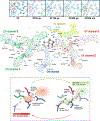
Figure 3.. The environment of the Mn cluster in PSII.
The Mn4CaOn cluster (Mn shown as blue, Ca as green, oxygen as red spheres) is embedded in a network of water channels (center, adapted from [9]) connecting it to the bulk. The main channels are labelled (‘O1 channel’, ‘O4 channel’ and ‘Cl1 channel’). Waters located in the room temperature X-ray structures are indicated by numbered circles and selected residues lining the channels are labeled. A pentamer of waters (W26-W30) located next to the O1 atom of the Mn cluster is indicated by a dashed circle. Changes in the electron density of this “water wheel” region at different time points in the S2 to S3 transition are clearly visible (top, adapted from [8]). These indicate high water mobility and a possible role of the water wheel and the O1 channel in water transport to the Mn cluster related to the water insertion event that takes place upon formation of the S3 state. A bottleneck in the Cl1 channel (highlighted by a dashed oval) changes configuration at around 150 μs into the S2 to S3 transition (bottom, adapted from [39]). This opening and closing of a potential “proton gate” could facilitate proton transfer from the cluster to the bulk during a specific time window in the S2 to S3 transition.

References

    1. Hillier W, and Messinger J (2005) Mechanism of photosynthetic oxygen production, in Photosystem II: The light-driven water:plastoquinone oxidoreductase, eds. Wydrzynski T, Satoh K, Springer.
    1. Shevela D, Kern JF, Govindjee G, Whitmarsh J, and Messinger J (2021) Photosystem II, in eLS, John Wiley & Sons, Ltd, pp. 1–16.
    1. Bergmann U, Kern J, Schoenlein RW, Wernet P, Yachandra VK, and Yano J (2021) Using X-ray free-electron lasers for spectroscopy of molecular catalysts and metalloenzymes. Nature Reviews Physics. - PMC - PubMed
    1. Margaritondo G, and Rebernik Ribic P (2011) A simplified description of X-ray free-electron lasers. J Synchrotron Radiat, 18 (Pt 2), 101–8. - PMC - PubMed
    1. Barty A, Caleman C, Aquila A, Timneanu N, Lomb L, White TA, Andreasson J, Arnlund D, Bajt S, Barends TRM, Barthelmess M, Bogan MJ, Bostedt C, Bozek JD, Coffee R, Coppola N, Davidsson J, DePonte DP, Doak RB, Ekeberg T, Elser V, Epp SW, Erk B, Fleckenstein H, Foucar L, Fromme P, Graafsma H, Gumprecht L, Hajdu J, Hampton CY, Hartmann R, Hartmann A, Hauser G, Hirsemann H, Holl P, Hunter MS, Johansson L, Kassemeyer S, Kimmel N, Kirian RA, Liang M, Maia FRNC, Malmerberg E, Marchesini S, Martin AV, Nass K, Neutze R, Reich C, Rolles D, Rudek B, Rudenko A, Scott H, Schlichting I, Schulz J, Seibert MM, Shoeman RL, Sierra RG, Soltau H, Spence JCH, Stellato F, Stern S, Strüder L, Ullrich J, Wang X, Weidenspointner G, Weierstall U, Wunderer CB, and Chapman HN (2012) Self-terminating diffraction gates femtosecond X-ray nanocrystallography measurements. Nature Photon, 6 (1), 35–40. - PMC - PubMed

Publication types