. 2022 Sep 17;11(10):e1689-e1694.
doi: 10.1016/j.eats.2022.06.003. eCollection 2022 Oct.

Anchor Arthropathy Caused by Cartilage Penetration: An Approach to Revision Hip Arthroscopy With Removal of Problematic Anchors

Affiliations

Anchor Arthropathy Caused by Cartilage Penetration: An Approach to Revision Hip Arthroscopy With Removal of Problematic Anchors

Payam W Sabetian et al. Arthrosc Tech. .

Abstract

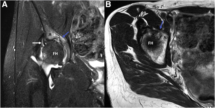
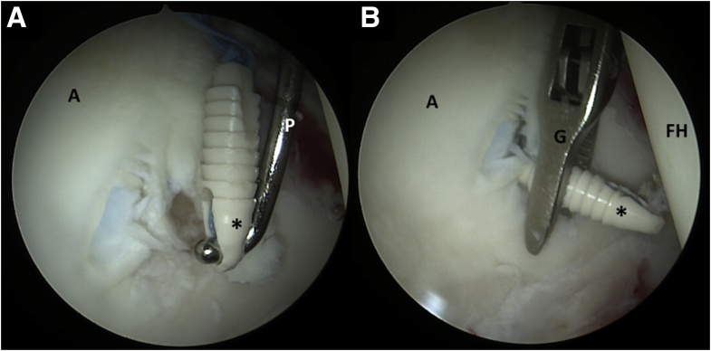
Hip arthroscopy has been proven to effectively treat labral tears in the setting of femoroacetabular impingement. Anchors used for this treatment have constantly evolved and improved to ensure safety and minimal invasion. However, acetabular drilling and anchor placement are technically challenging due to the concavity of the acetabular articular surface, limited angles for anchor insertion, and finite bone availability in the anterior and posterior column. Inadequate technique can result in protruding anchors, which may lead to full-thickness articular cartilage damage, manifesting in pain, mechanical symptoms, and impaired function. This Technical Note demonstrates arthroscopic removal of protruding anchors and management of the iatrogenic grade IV cartilage damage. In this description, the technical pearls and pitfalls of acetabular anchor placement to treat labral pathology are presented along with the aforementioned technique.


Figures

Fig 1
Fig 1
(A) Coronal T2 image of a fat-saturated magnetic resonance arthrogram (MRA) of the right hip, where a labral tear can be visualized (white arrow). (B) Axial T2 image of a fat-saturated MRA of the right hip. Hyperintense images produced by the protruding anchors can be seen in both views, disrupting articular cartilage of the acetabulum (blue arrow). (A, acetabulum; FH, femoral head.)
Fig 2
Fig 2
(A) The patient is placed in the modified supine position and the anterior inferior iliac spine is marked (∗). (B) The right hip is shown, with patient’s head to the left and feet to the right. The 4 portals used are identified: anterolateral (AL), mid-anterior (MA), distal anterolateral accessory (DALA), and posterolateral (PL). (This figure was previously published by Sabetian et al. under the terms of https://creativecommons.org/licenses/by-nc-nd/4.0/.)
Fig 3
Fig 3
Intraoperative images during a revision diagnostic arthroscopy visualized with a 70° arthroscope from the anterolateral (AL) portal, assessing the articular surface of the acetabulum (A) and femoral head (FH), as well as the labrum (L). The labrum is assessed with a probe (P) introduced through the mid-anterior portal, finding a combined Seldes 1 and 2 tear (blue arrow). Anchors used to repair the labrum on previous surgery are visualized protruding through the acetabular surface (black arrow), which results in chondral damage to the femoral head is as signaled with the white arrow.
Fig 4
Fig 4
Intraoperative images during a revision diagnostic arthroscopy with the patient in a supine position, visualized through the anterolateral (AL) portal. (A) Anchors used during previous surgery to repair the labrum are evidenced protruding and causing anchor arthropathy (blue arrows). (B) Arthroscopic image showing articular cartilage damage caused by the protruding anchors are further uncovered with the use of a probe (black arrow). (C) Extensive chondral damage through subchondral bone is evidenced after removal of the protruding anchors (black star); intact acetabular cartilage is marked with a blue star. (D) Damage caused by protruding anchors has extended to the articular surface of the femoral head (identified with a black star), surrounded by undamaged cartilage (blue star). (A, acetabulum; FH, femoral head.)
Fig 5
Fig 5
Intraoperative images during removal of protruding anchors visualized with a 70° arthroscope from the anterolateral (AL) portal. (B) An arthroscopic probe (P) is introduced through the midanterior portal and used to expose and mobilize anchors used for labral repair in a previous procedure, which have protruded through articular cartilage (black star). (B) An arthroscopic grasper (G) is used to complete the en bloc removal of the anchors. (A, acetabulum; FH, femoral head.)

References

    1. Ferguson S.J., Bryant J.T., Ganz R., Ito K. The influence of the acetabular labrum on hip joint cartilage consolidation: a poroelastic finite element model. J Biomech. 2000;33:953–960. - PubMed
    1. Nepple J.J., Philippon M.J., Campbell K.J., et al. The hip fluid seal—Part II: The effect of an acetabular labral tear, repair, resection, and reconstruction on hip stability to distraction. Knee Surg Sports Traumatol Arthrosc. 2014;22:730–736. - PubMed
    1. Maldonado D.R., Kyin C., Chen S.L., et al. In search of labral restoration function with hip arthroscopy: outcomes of hip labral reconstruction versus labral repair: A systematic review. HIP Int. 2021;31:704–713. - PubMed
    1. Kyin C., Maldonado D.R., Go C.C., Shapira J., Lall A.C., Domb B.G. Mid- to long-term outcomes of hip arthroscopy: A systematic review. Arthroscopy. 2021;37:1011–1025. - PubMed
    1. Schüttler K.F., Schramm R., El-Zayat B.F., Schofer M.D., Efe T., Heyse T.J. The effect of surgeon’s learning curve: Complications and outcome after hip arthroscopy. Arch Orthop Trauma Surg. 2018;138:1415–1421. - PubMed

LinkOut - more resources