Review
. 2022 Oct 13:10:1017305.
doi: 10.3389/fchem.2022.1017305. eCollection 2022.

A review of cardiac troponin I detection by surface enhanced Raman spectroscopy: Under the spotlight of point-of-care testing

Affiliations
Review

A review of cardiac troponin I detection by surface enhanced Raman spectroscopy: Under the spotlight of point-of-care testing

Anel I Saviñon-Flores et al. Front Chem. .

Abstract

Cardiac troponin I (cTnI) is a biomarker widely related to acute myocardial infarction (AMI), one of the leading causes of death around the world. Point-of-care testing (POCT) of cTnI not only demands a short turnaround time for its detection but the highest accuracy levels to set expeditious and adequate clinical decisions. The analytical technique Surface-enhanced Raman spectroscopy (SERS) possesses several properties that tailor to the POCT format, such as its flexibility to couple with rapid assay platforms like microfluidics and paper-based immunoassays. Here, we analyze the strategies used for the detection of cTnI by SERS considering POCT requirements. From the detection ranges reported in the reviewed literature, we suggest the diseases other than AMI that could be diagnosed with this technique. For this, a section with information about cardiac and non-cardiac diseases with cTnI release, including their release kinetics or cut-off values are presented. Likewise, POCT features, the use of SERS as a POCT technique, and the biochemistry of cTnI are discussed. The information provided in this review allowed the identification of strengths and lacks of the available SERS-based point-of-care tests for cTnI and the disclosing of requirements for future assays design.

Keywords: SERS-based immunoassays; acute myocardial infarction (AMI); cardiac troponin I (cTnI); point-of-care testing; surface-enhanced Raman spectroscopy (SERS).


Conflict of interest statement

The authors declare that the research was conducted in the absence of any commercial or financial relationships that could be construed as a potential conflict of interest.

Figures
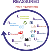
FIGURE 1
FIGURE 1
Meaning of REASSURED acronym.
FIGURE 2
FIGURE 2
Release kinetics of cardiac and non-cardiac diseases: (A) AMI, myocarditis, and Chronic Heart Failure (CHF), adapted from Mahajan and Jarolim (2011); Abdolrahim et al. (2015), and (B) Arterial Hypertension (AH), Acute Pulmonary Embolism (APE), and Strenuous Exercise (SE), adapted from Douketis et al. (2002); Taddei et al. (2011); Brzezinski et al. (2019).
FIGURE 3
FIGURE 3
Epitopic map of cTnI containing the amino acid sequences of the most important external and internal epitopes adapted from HyTest (2022) and Filatov et al. (1998).
FIGURE 4
FIGURE 4
SERS measurement routes. (A) in solution, (B,C) on substrates.
FIGURE 5
FIGURE 5
Schematic representation of the aptasensor based on a flower-shaped silver magnetic nanocomposite for the label-free detection of cTnI by SERS, adapted from Alves et al. (2020).
FIGURE 6
FIGURE 6
Schematic representation of different cTnI detection systems working in solution. (A) sandwich immunoassay, adapted from Hu et al. (2021) and (B) 3D AuNPs-PEN substrate, adapted from Lee et al. (2022).
FIGURE 7
FIGURE 7
SERS-based competitive immunoassay proposal by (A) Garza and Cote (2017) under the principle of sequential saturation, adapted from Garza and Cote (2017), homemade collection device used for the SERS experiments in incise (B).
FIGURE 8
FIGURE 8
Schematic representation of SERS-based sandwich immunoassay for the independent quantification of cTnI and CK-MB, adapted from Cheng et al. (2019).
FIGURE 9
FIGURE 9
Schematic representation of aptamer-based SERS assays for cTnI: (A) the sandwich assay using an atomically flat Au nanoplate as the plasmonic substrate, adapted from Lee et al. (2020), (B) assay using the Bradford method, adapted from Lin et al. (2021b).
FIGURE 10
FIGURE 10
Schematic representation of the sandwich immunoassay integrated in an active microfluidic device for the quantification of cTnI and neuropeptide Y, adapted from Wen et al. (2020).
FIGURE 11
FIGURE 11
Schematic representation of the multiplex SERS-based lateral flow assay proposed by Zhang et al. (2018a).
FIGURE 12
FIGURE 12
(A) The four different types of SERS substrate used to elaborate the capture probes in the SERS-based LFIA strip presented by Bai et al. (2018), (B) Schematic representation of the aptamer-based SERS assay on paper platform, adapted from Tu et al. (2020).

References

    1. Abdolrahim M., Rabiee M., Alhosseini S. N., Tahriri M., Yazdanpanah S., Tayebi L. (2015). Development of optical biosensor technologies for cardiac troponin recognition. Anal. Biochem. 485, 1–10. 10.1016/J.AB.2015.06.003 - DOI - PubMed
    1. Abel G. (2015). Current status and future prospects of point-of-care testing around the globe. Expert Rev. Mol. diagn. 15 (7), 853–855. 10.1586/14737159.2015.1060126 - DOI - PubMed
    1. Aboughdir M., Kirwin T., Abdul Khader A., Wang B. (2020). Prognostic value of cardiovascular biomarkers in COVID-19: A review. Viruses 12 (5), 527. 10.3390/v12050527 - DOI - PMC - PubMed
    1. Al-Hillawi E., Bhandari D. G., Trayer H. R., Trayer I. P. (1995). The effects of phosphorylation of cardiac troponin-I on its interactions with actin and cardiac troponin-C. Eur. J. Biochem. 228 (3), 962–970. 10.1111/J.1432-1033.1995.0962M.X - DOI - PubMed
    1. Alessandri I., Lombardi J. R. (2020). Editorial: Surface enhanced Raman scattering: New theoretical approaches, materials and strategies. Front. Chem. 8, 63. 10.3389/fchem.2020.00063 - DOI - PMC - PubMed