. 2022 Oct 14:13:983000.
doi: 10.3389/fimmu.2022.983000. eCollection 2022.

mRNA vaccine with unmodified uridine induces robust type I interferon-dependent anti-tumor immunity in a melanoma model

Affiliations

mRNA vaccine with unmodified uridine induces robust type I interferon-dependent anti-tumor immunity in a melanoma model

Chutamath Sittplangkoon et al. Front Immunol. .

Abstract

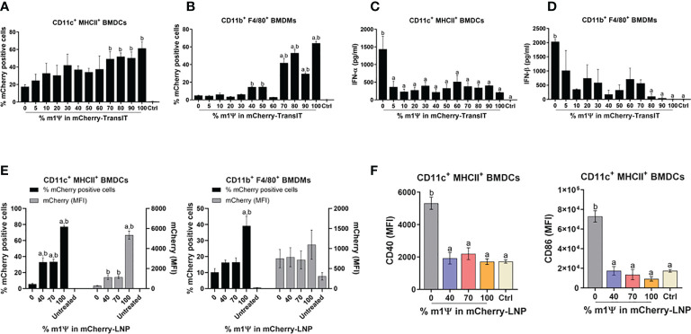
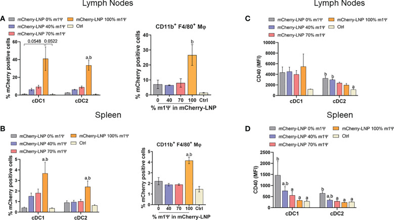
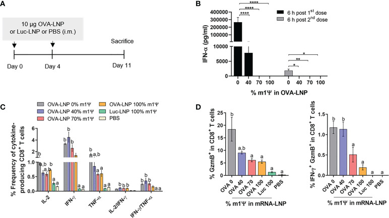
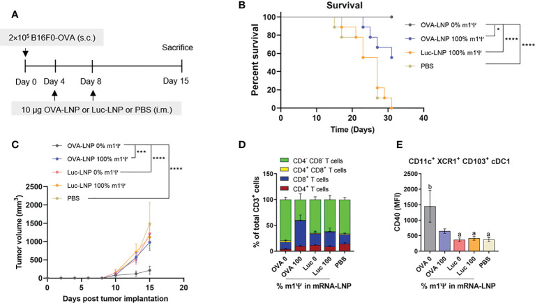
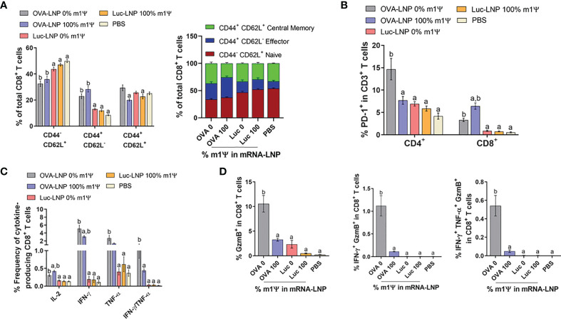
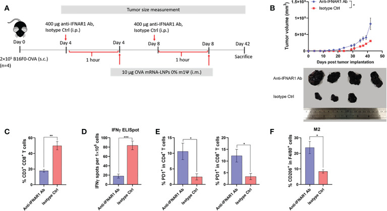
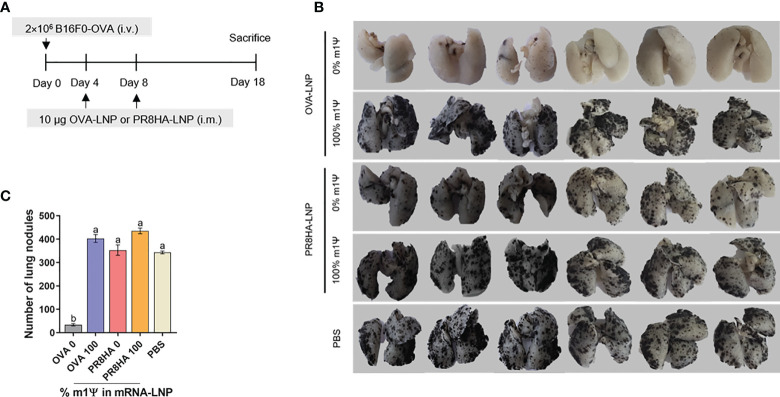
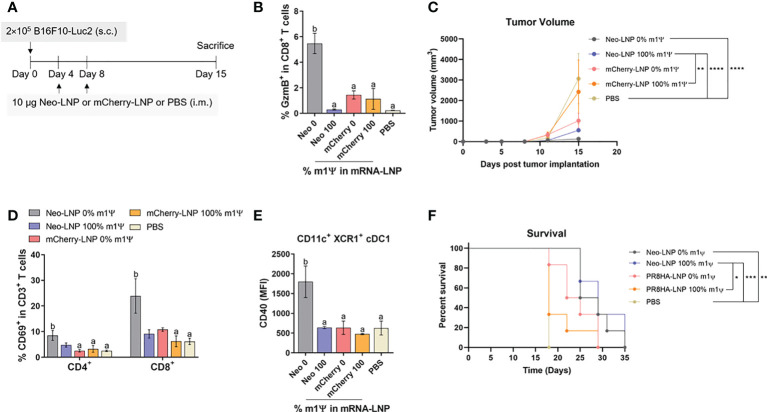
An mRNA with unmodified nucleosides induces type I interferons (IFN-I) through the stimulation of innate immune sensors. Whether IFN-I induced by mRNA vaccine is crucial for anti-tumor immune response remains to be elucidated. In this study, we investigated the immunogenicity and anti-tumor responses of mRNA encoding tumor antigens with different degrees of N1-methylpseudouridine (m1Ψ) modification in B16 melanoma model. Our results demonstrated that ovalbumin (OVA) encoding mRNA formulated in a lipid nanoparticle (OVA-LNP) induced substantial IFN-I production and the maturation of dendritic cells (DCs) with negative correlation with increasing percentages of m1Ψ modification. In B16-OVA murine melanoma model, unmodified OVA-LNP significantly reduced tumor growth and prolonged survival, compared to OVA-LNP with m1Ψ modification. This robust anti-tumor effect correlated with the increase in intratumoral CD40+ DCs and the frequency of granzyme B+/IFN-γ+/TNF-α+ polyfunctional OVA peptide-specific CD8+ T cells. Blocking type I IFN receptor completely reversed the anti-tumor immunity of unmodified mRNA-OVA reflected in a significant decrease in OVA-specific IFN-γ secreting T cells and enrichment of PD-1+ tumor-infiltrating T cells. The robust anti-tumor effect of unmodified OVA-LNP was also observed in the lung metastatic tumor model. Finally, this mRNA vaccine was tested using B16 melanoma neoantigens (Pbk-Actn4) which resulted in delayed tumor growth. Taken together, our findings demonstrated that an unmodified mRNA vaccine induces IFN-I production or the downstream signaling cascades which plays a crucial role in inducing robust anti-tumor T cell response for controlling tumor growth and metastasis.

Keywords: cancer immunotherapy; mRNA vaccine; melanomas; type I interferon; unmodified nucleosides.


Conflict of interest statement

PL and YT are employees of Acuitas Therapeutics, a company involved in the development of mRNA-LNP therapeutics. YT, DW, and M-GA are named on patents that describe lipid nanoparticles for delivery of nucleic acid therapeutics, including mRNA and the use of modified mRNA in lipid nanoparticles as a vaccine platform. The remaining authors declare that the research was conducted in the absence of any commercial or financial relationships that could be construed as a potential conflict of interest.

Figures

Figure 1
Figure 1
TransIT and LNP efficiently delivered mRNA to murine BMDCs and BMDMs in vitro. (A–D) mCherry-encoding mRNA modified with different % of m1Ψ (0.1 µg) were transfected into BMDCs and BMDMs using TransIT reagent. The frequencies of mCherry positive (A) BMDCs and (B) BMDMs were determined by flow cytometry at 48 hr after transfection. (C) The levels of IFN-α and (D) IFN-β released upon mRNA transfection from BMDCs and BMDMs, respectively were examined by ELISA. (E) mCherry-encoding mRNA modified with different % of m1Ψ (0.1 µg) were delivered into BMDCs and BMDMs using LNP. The frequencies of mCherry positive BMDCs (left) and BMDMs (right) and MFI of mCherry delivered by LNP were determined by flow cytometry. (F) MFI of CD40 (left) and CD86 (right) on BMDCs upon mRNA-LNP transfection was shown. The control were untranfected cells. The results are presented as the mean ± SEM of at least duplicate samples and experiments were performed at least two times. Statistical significance by one-way ANOVA with Bonferroni multiple comparisons test were indicated when p < 0.05 compared to the unmodified target antigen: a, or control (PBS): b.
Figure 2
Figure 2
mRNA-LNP uptake by APCs in lymph nodes (LNs) and spleens in vivo. Mice were intramuscular injected with 10 µg of mCherry mRNA-LNP with 0, 40, 70, or 100% of m1Ψ modification and the control group received PBS. At 48 hr of mRNA administration, quantification of mCherry-positive cells in (A) LNs and (B) spleen of conventional type 1 (cDC1) and conventional type 2 (cDC2) dendritic cells (left), and MΦ (right) were determined. MFI of costimulatory molecule CD40 on cDC1 and cDC2 from (C) LNs and (D) spleen were shown. The results are presented as the mean ± SEM of biologically independent mice (n = 6) per group. Statistical significance by one-way ANOVA with Bonferroni multiple comparisons test when p < 0.05 compared to the unmodified target antigen: a, or control (PBS): (D) cDC1 subset was defined as Dump- (B220- NK1.1- CD3- TER-119- CD19-) CD11c+ MHCII+ XCR1+. cDC2 subset was defined as Dump- (B220- NK1.1- CD3- TER-119- CD19-) CD11c+ MHCII+ CD172a+.
Figure 3
Figure 3
Immunization of mRNA-LNP elicits robust antigen-specific CD8+ T cell responses. (A) Schematic representation of the immunization regimen (see methods for details). (B) IFN-α concentration in the serum 6 hr after the first (day0) and the second (day4) immunizations of OVA mRNA-LNP were detected by ELISA. (C, D) The frequencies of IL-2, IFNγ, TNFα and Granzyme B (GzmB) and IFN-γ/GzmB-producing CD8+ T cells after 6 hr stimulation with OVA257-264 (SIINFEKL) were measured by flow cytometry. The results are presented as the mean ± SEM, n = 4-5 biologically independent mice per group. Statistical significance: Statistical significance by one-way ANOVA with Bonferroni multiple comparisons test were indicated as (*)p < 0.05, (**)p < 0.01 and (****)p < 0.0001 or p < 0.05 compared to the unmodified target antigen: a, or control (PBS): b.
Figure 4
Figure 4
Intramuscular immunization of unmodified OVA-LNP inhibits tumor growth and prolongs survival. (A) Schematic representation of the immunization and tumor implantatioin schedule. (B) The percentages of survival mice was followed until day 31 after tumor implantation. Mice that reached the maximal allowed tumor size of 20 mm, or 400 mm2 were euthanized and recorded as having tumor areas of 400 mm2 (n = 9). (C) Tumor volume was shown during the 15 days after tumor implantation (n = 10). (D) Compositions of tumor-infiltrating T cells and (E) MFI of costimulatory molecule CD40 on cDC1 are shown (n = 6). The results are presented as the mean ± SEM. Statistical significance: (*)p < 0.05, (***)p < 0.001 and (****)p < 0.0001 by two-way ANOVA with Bonferroni multiple comparisons test. Survival curves were compared using log-rank (Mantel–Cox) test.
Figure 5
Figure 5
Immunization of unmodified OVA-LNP enhances the activation of antigen-specific CD8+ T cells. Mice were treated as described in Figure 4A and the splenocytes were re-stimulated for 6 h with OVA257-264 (SIINFEKL) peptide. (A) The frequencies of naïve cells (CD44-CD62L+), effector cells (CD44+CD62L-), and memory cells (CD44+CD62L+) in CD3+ CD8+ T cell subsets (B) the frequencies of PD-1+ CD4+ and CD8+ T cells and (C) the frequencies of cytokines-producing CD8+ T cells are shown. (D) The OVA257-264-specifc responses were determined and the percentages of CD8+ T cells producing GzmB, IFN-γ/GzmB, or IFN-γ/TNF-α/GzmB are shown. The results are presented as the mean ± SEM of biologically independent mice (n=7) per group. Statistical significance by one-way ANOVA with Bonferroni multiple comparisons test when p < 0.05 compared to the unmodified target antigen: a, control (PBS): b.
Figure 6
Figure 6
Type I IFN signaling promotes antitumor effect and modulated immune cell profile. (A) Scheme of immunization regimen and IFNAR1 antibody or isotype control treatment. See details in methods. (B) The tumor volume (top panel) and the representative tumor mass (lower panel) harvested 42 days after the tumor implantation from mice treated with anti-IFNAR1 antibody or isotype control followed by immunization with unmodified OVA-LNP. (C) The frequency of splenic CD8+ T cells was examined by flow cytometry. (D) ELISpot of IFN-γ producing cells among splenocytes after 48 hr of ex vivo re-stimulation with OVA on day 42 after tumor implantation and mRNA vaccine treatments is shown. (E) The frequency of PD-1+ cells among tumor infiltrated CD4+ (left) and CD8+ (right) T cells were examined by flow cytometry. (F) The frequency of tumor infiltrated CD206+ macrophages was determined by flow cytometry. The results are presented as the mean ± SEM, n = 4 biologically independent mice per group. Statistical significance: (*)p < 0.05, (**)p < 0.01, (***)p < 0.001 by unpaired two-tailed Student’s t test.
Figure 7
Figure 7
Immunization of unmodified mRNA-LNP inhibits lung metastasis of B16F0-OVA melanoma. (A) Schematic immunization schedule for melanoma metastatic model. See methods for details. (B) The lungs were observed and (C) the metastatic nodules on the surface of the lungs were counted. The results are presented as the mean ± SEM of biologically independent mice (n = 6) per group. Statistical significance by one-way ANOVA with Bonferroni multiple comparisons test when p < 0.05 compared to the unmodified target antigen: a, control: b.
Figure 8
Figure 8
Neoantigens (Pbk-Actn4) encoding mRNA-LNP inhibits tumor growth in vivo and prolongs survival. (A) Schematic representation of the immunization regimen to test the anti-tumor efficacy of neoantigen encoding mRNA-LNP. See methods for details. (B) The frequency of GzmB-producing splenic CD8+ T cells on CD3+ CD8+ T cell subsets was determined by flow cytometry. (C) Tumor volume was measured (n = 10) (D) The frequencies of CD69+ T cells among tumor infiltrated CD4+ and CD8+ T cells are shown. (E) The MFI of costimulatory molecule CD40 on tumor-infiltrating cDC1 is shown (n = 4). (F) The percentage of survival was followed until day 35 (n = 6). GzmB, granzyme; cDC1, conventional type 1 dendritic cell. (B) The results are presented as the mean ± SEM. Statistical significance: (*)p < 0.05, (**)p < 0.01, (***)p < 0.001 and (****)p < 0.0001 by two-way ANOVA with Bonferroni multiple comparisons test. Survival curves were compared using log-rank (Mantel–Cox) test.

References

    1. Wang RF, Wang HY. Immune targets and neoantigens for cancer immunotherapy and precision medicine. Cell Res (2017) 27(1):11–37. doi: 10.1038/cr.2016.155 - DOI - PMC - PubMed
    1. Li W, Joshi MD, Singhania S, Ramsey KH, Murthy AK. Peptide vaccine: Progress and challenges. Vaccines (Basel) (2014) 2(3):515–36. doi: 10.3390/vaccines2030515 - DOI - PMC - PubMed
    1. Zhang L, Huang Y, Lindstrom AR, Lin TY, Lam KS, Li Y. Peptide-based materials for cancer immunotherapy. Theranostics (2019) 9(25):7807–25. doi: 10.7150/thno.37194 - DOI - PMC - PubMed
    1. Nelde A, Rammensee HG, Walz JS. The peptide vaccine of the future. Mol Cell Proteom (2021) 20:100022. doi: 10.1074/mcp.R120.002309 - DOI - PMC - PubMed
    1. Lopes A, Vandermeulen G, Preat V. Cancer DNA vaccines: current preclinical and clinical developments and future perspectives. J Exp Clin Cancer Res (2019) 38(1):146. doi: 10.1186/s13046-019-1154-7 - DOI - PMC - PubMed

Publication types