. 2022 Oct 13:13:1042463.
doi: 10.3389/fimmu.2022.1042463. eCollection 2022.

DNA-PKcs restricts Zika virus spreading and is required for effective antiviral response

Affiliations

DNA-PKcs restricts Zika virus spreading and is required for effective antiviral response

Daniel de Oliveira Patricio et al. Front Immunol. .

Abstract

Zika virus (ZIKV) is a single-strand RNA mosquito-borne flavivirus with significant public health impact. ZIKV infection induces double-strand DNA breaks (DSBs) in human neural progenitor cells that may contribute to severe neuronal manifestations in newborns. The DNA-PK complex plays a critical role in repairing DSBs and in the innate immune response to infection. It is unknown, however, whether DNA-PK regulates ZIKV infection. Here we investigated the role of DNA-PKcs, the catalytic subunit of DNA-PK, during ZIKV infection. We demonstrate that DNA-PKcs restricts the spread of ZIKV infection in human epithelial cells. Increased ZIKV replication and spread in DNA-PKcs deficient cells is related to a notable decrease in transcription of type I and III interferons as well as IFIT1, IFIT2, and IL6. This was shown to be independent of IRF1, IRF3, or p65, canonical transcription factors necessary for activation of both type I and III interferon promoters. The mechanism of DNA-PKcs to restrict ZIKV infection is independent of DSB. Thus, these data suggest a non-canonical role for DNA-PK during Zika virus infection, acting downstream of IFNs transcription factors for an efficient antiviral immune response.

Keywords: DNA-PKcs; Zika virus; double-strand DNA breaks; infection; interferon.


Conflict of interest statement

The authors declare that the research was conducted in the absence of any commercial or financial relationships that could be construed as a potential conflict of interest.

Figures
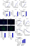
Figure 1
Figure 1
DNA-PKcs is critical for control of ZIKV infection. Virus replication measured by plaque assay, expressed as plaque-forming units per mL (PFU/mL), on (A) A549WT and A549 PRKDC-/- or (B) RPEWT and RPE PRKDC-/- cells infected with ZIKV at indicated m.o.i. and time. RT-qPCR analysis to measure ZIKV RNA in (C) A549WT and A549 PRKDC-/- or (D) RPEWT and RPE PRKDC-/- cells infected at indicated m.o.i. and time. (E) A549WT and A549 PRKDC-/- or (F) RPEWT and RPE PRKDC-/- cells infected with 50 PFU of ZIKV at 48 hours in semi-solid medium, then ZIKV-E protein (green) was stained for immunofluorescence analysis, and the relative area of infection percentage was measured using the ImageJ software. The cell nuclei were stained with DAPI (blue). (G) Percentage of ZIKV-infected A549WT and A549 PRKDC-/- cells at indicated m.o.i. and time, analyzed by flow cytometry. (H) Viability analysis by MTT assay of ZIKV-infected A549WT and A549 PRKDC-/- cells relative to uninfected cells (mock) at indicated m.o.i. and time. (I) A549WT and A549 PRKDC-/- were pretreated with NU7441 (0.5 and 1 µM) at 24 hours followed by ZIKV infection (m.o.i. 1) at 24 hours. We used two-way ANOVA with Sidak’s correction in (A–D, G, H), and unpaired two-tailed Student’s t-test was used in (E, F). * p<0.05, n = 3, error bars ± SEM.
Figure 2
Figure 2
ZIKV infection does not induce DSB in A549 cells. A549WT, A549 PRKDC-/- and 1 µM NU7441 pre-treated A549WT infected with ZIKV (m.o.i. 1) at 24 hours. Stimulation with 3 µM etoposide for 12 hours was used as a DSB positive control. (A) Immunofluorescence to analyze γH2AX (green) in the ZIKV-infected cells (red, ZIKV-E protein). The cell nuclei were stained with DAPI (blue). (B) Percentage of γH2AX foci per cell showed in (A). *Compared with WT cells; #Compared with mock. We used two-way ANOVA with Sidak’s correction. * or # p<0.05, n = 3, error bars ± SEM.
Figure 3
Figure 3
DNA-PKcs regulates interferon-related genes during ZIKV infection. (A) RT-qPCR to measure the expression of mRNA for indicated genes on A549WT and A549RIGI-/- cells infected with ZIKV m.o.i. 1 for 24 hours. (B) RT-qPCR to measure the expression of mRNA for indicated genes on A549WT and A549 PRKDC-/- cells infected with ZIKV m.o.i. 0.1 at the indicated time or (C) m.o.i. 1 at 24 hours. (D) Expression of mRNA for NFKBIA and IL6, determined by RT-qPCR on A549WT and A549 PRKDC-/- cells infected with ZIKV m.o.i. 1 at 24 hours. We used unpaired two-tailed Student’s t-test in (A, C, D), and two-way ANOVA with Sidak’s correction in (B). ND: Non-detected. * p<0.05, n = 3, error bars ± SEM.
Figure 4
Figure 4
ZIKV induces IRF1, IRF3, and p65 nuclei accumulation independent of DNA-PKcs. Immunofluorescence analysis for localization of endogenous (A) IRF3 (red) and (B) IRF1 (red) on A549WT and A549RIGI-/- cells infected with ZIKV m.o.i. 1 (green, ZIKV-E protein) at 36 hours (left panel), and quantified by scoring cells with nuclear staining (right panel, n = 3, counts of at least 30 nuclei per slide). (C–E) Immunofluorescence analysis for localization of endogenous (C) IRF3 (red), (D) IRF1 (red), and (E) p65 (red) on A549WT and A549 PRKDC-/- cells infected with ZIKV m.o.i. 1 (green, ZIKV-E protein) at 24 hours (left panel), and quantified by scoring cells with nuclear staining (right panel, n = 3, counts of at least 30 nuclei per slide). TNF was used as a positive control in (E). Cell nuclei were stained with DAPI (grey). We used unpaired two-tailed Student’s t-test. N = 3, error bars ± SEM. ND: non-detected.

References

    1. Masmejan S, Musso D, Vouga M, Pomar L, Dashraath P, Stojanov M, et al. . Zika virus. Pathogens (2020) 9:898. doi: 10.3390/pathogens9110898 - DOI - PMC - PubMed
    1. Cardoso CW, Paploski IAD, Kikuti M, Rodrigues MS, Silva MMO, Campos GS, et al. . Outbreak of exanthematous illness associated with zika, chikungunya, and dengue viruses, Salvador, Brazil. Emerg Infect Dis (2015) 21:2274–6. doi: 10.3201/eid2112.151167 - DOI - PMC - PubMed
    1. Musso D, Nilles EJ, Cao-Lormeau V-M. Rapid spread of emerging zika virus in the pacific area. Clin Microbiol Infection (2014) 20:O595–6. doi: 10.1111/1469-0691.12707 - DOI - PubMed
    1. Duffy MR, Chen T-H, Hancock WT, Powers AM, Kool JL, Lanciotti RS, et al. . Zika virus outbreak on yap island, federated states of Micronesia. New Engl J Med (2009) 360:2536–43. doi: 10.1056/NEJMoa0805715 - DOI - PubMed
    1. Dick GWA, Kitchen SF, Haddow AJ. Zika virus (I). isolations and serological specificity. Trans R Soc Trop Med Hyg (1952) 46:509–20. doi: 10.1016/0035-9203(52)90042-4 - DOI - PubMed

Publication types