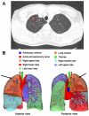
Three-dimensional computed tomography reconstruction in video-assisted thoracoscopic segmentectomy (DRIVATS): A prospective, multicenter randomized controlled trial
- PMID: 36311929
- PMCID: PMC9606583
- DOI: 10.3389/fsurg.2022.941582
Three-dimensional computed tomography reconstruction in video-assisted thoracoscopic segmentectomy (DRIVATS): A prospective, multicenter randomized controlled trial
Abstract
Objective: Anatomical segmentectomy has been proven to be a viable surgical treatment for small-size peripheral lung nodules. Three-dimensional (3D) reconstruction computed tomography (CT) has been proposed as an effective approach to overcome the challenges of encountering pulmonary anatomical variations when performing segmentectomy. Therefore, to further investigate the usefulness of preoperative 3D reconstruction CT in segmentectomy, we will conduct this prospective, multicenter randomized controlled DRIVATS study to compare the use of 3D reconstruction CT with standard chest CT in video-assisted segmentectomy (ClinicalTrials.gov ID: NCT04004494).
Methods: This study began in July 2019 and a total of 190 patients will be accrued from three clinical centers within 4 years. The main inclusion criteria are patients with a single peripheral nodule 0.8-2 cm with at least one of the following requirements: (i) histology of adenocarcinoma in situ; (ii) nodule has ≥50% ground-glass appearance on CT; (iii) radiologic surveillance confirms a long doubling time (≥400 days). Surgical procedures include segmental resection of the lesion and mediastinal lymph node sampling (subsegmental resection or combined subsegmental resection will not be included in this study). The primary endpoint is operative time. The secondary endpoints include incidence of change of surgical plan, intraoperative blood loss, conversion rate, operative accident event, incidence of postoperative complications, postoperative hospital stay, length of hospitalization, duration of chest tube placement, postoperative 30-day mortality, dissection of lymph nodes, overall survival, disease-free survival, preoperative lung function, and postoperative lung function.
Discussion: This multicenter DRIVATS study aims to verify the usefulness of preoperative 3D reconstruction CT compared with standard chest CT in segmentectomy. If successfully completed, this multicenter prospective study will provide a higher level of evidence for the use of 3D reconstruction CT in segmentectomy.
Keywords: 3D reconstruction CT; multicenter randomized controlled trial; pulmonary nodules; segmentectomy; video-assisted thoracoscopic.
© 2022 Niu, Chen, Jin, Zheng, Gong, Nie, Jiang, Zhong, Chen and Li.
Conflict of interest statement
The authors declare that the research was conducted in the absence of any commercial or financial relationships that could be construed as a potential conflict of interest.
Figures


Similar articles
-
Three-dimensional reconstruction computed tomography in thoracoscopic segmentectomy: a randomized controlled trial.Eur J Cardiothorac Surg. 2024 Jul 1;66(1):ezae250. doi: 10.1093/ejcts/ezae250. Eur J Cardiothorac Surg. 2024. PMID: 38936342 Clinical Trial.
-
The advantages of preoperative 3D reconstruction over 2D-CT in thoracoscopic segmentectomy.Updates Surg. 2024 Dec;76(8):2875-2883. doi: 10.1007/s13304-024-01965-6. Epub 2024 Sep 29. Updates Surg. 2024. PMID: 39342519 Free PMC article.
-
Preoperative 3-dimensional computed tomography lung simulation before video-assisted thoracoscopic anatomic segmentectomy for ground glass opacity in lung.J Thorac Dis. 2018 Dec;10(12):6598-6605. doi: 10.21037/jtd.2018.10.126. J Thorac Dis. 2018. PMID: 30746205 Free PMC article.
-
Novel techniques for video-assisted thoracoscopic surgery segmentectomy.J Thorac Dis. 2018 Jun;10(Suppl 14):S1671-S1676. doi: 10.21037/jtd.2018.05.207. J Thorac Dis. 2018. PMID: 30034834 Free PMC article. Review.
-
In patients with resectable non-small-cell lung cancer, is video-assisted thoracoscopic segmentectomy a suitable alternative to thoracotomy and segmentectomy in terms of morbidity and equivalence of resection?Interact Cardiovasc Thorac Surg. 2014 Jul;19(1):107-10. doi: 10.1093/icvts/ivu080. Epub 2014 Apr 10. Interact Cardiovasc Thorac Surg. 2014. PMID: 24722517 Review.
Cited by
-
Three-Dimensional Imaging-Guided Lung Anatomic Segmentectomy: A Single-Center Preliminary Experiment.Medicina (Kaunas). 2023 Nov 26;59(12):2079. doi: 10.3390/medicina59122079. Medicina (Kaunas). 2023. PMID: 38138182 Free PMC article.
-
Anatomy of the lung revisited by 3D-CT imaging.Video Assist Thorac Surg. 2023 Jun 30;8:17. doi: 10.21037/vats-23-21. Epub 2023 May 24. Video Assist Thorac Surg. 2023. PMID: 37711275 Free PMC article.
-
Harnessing 3D-CT Simulation and Planning for Enhanced Precision Surgery: A Review of Applications and Advancements in Lung Cancer Treatment.Cancers (Basel). 2023 Nov 14;15(22):5400. doi: 10.3390/cancers15225400. Cancers (Basel). 2023. PMID: 38001660 Free PMC article. Review.
-
Effect of 18F-FDG PET/CT Imaging Combined with 3D-Printed Templates on Biopsy Efficacy in Patients with Lung Tumor.Pak J Med Sci. 2025 Apr;41(4):1047-1051. doi: 10.12669/pjms.41.4.9828. Pak J Med Sci. 2025. PMID: 40290235 Free PMC article.
References
Associated data
LinkOut - more resources
Full Text Sources
Medical

