

Cervical subtotal discectomy prosthesis validated in non-human primate model: A novel artificial cervical disc replacement concept?
- PMID: 36312530
- PMCID: PMC9606661
- DOI: 10.3389/fbioe.2022.997877
Cervical subtotal discectomy prosthesis validated in non-human primate model: A novel artificial cervical disc replacement concept?
Abstract
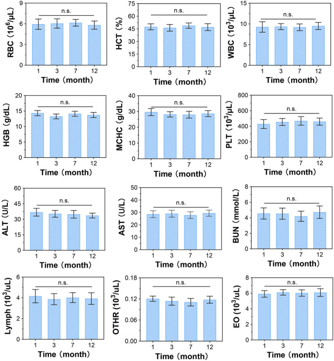
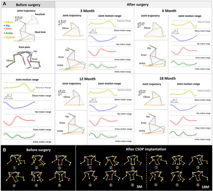
Objective: To evaluate the biological function of cervical subtotal discectomy prosthesis (CSDP) implantation in a non-human primate model. Methods: A CSDP was tested for cytocompatibility and osseointegration capacity before implantation in non-human primates. Subsequently, the CSDP was improved based on three-dimensional CT measurements of the non-human primate cervical spine. Eight cynomolgus monkeys were selected for removal of the intervertebral disc and lower endplate of the C5/6 segment to complete the model construction for CSDP implantation. In 18-month follow-up, physiological indices, radiology, and kinematics were assessed to estimate the biological function of the CSDP in non-human primates, including biosafety, osseointegration, and biomechanics. Results: Co-cultured with the CSDP constituent titanium alloy (Ti6Al4V-AO), the mouse embryo osteoblast precursor cell MC3T3-E1 obtained extended adhesion, remarkable viability status, and cell proliferation. After implantation in the mouse femur for 28 days, the surface of Ti6Al4V-AO was covered by a large amount of new cancellous bone, which formed further connections with the femur cortical bone, and no toxicity was detected by blood physiology indices or histopathology. After completing implantation in primate models, no infection or osteolysis was observed, nor was any subsidence or displacement of the CSDP observed in CT scans in the 18-month follow-up. In particular, the interior of the cervical vertebra fixation structure was gradually filled with new trabecular bone, and the CSDP had achieved fixation and bony fusion in the vertebral body at 1 year post-operation. Meanwhile, no signs of inflammation, spinal cord compression, adjacent segment degeneration, or force line changes were observed in subsequent MRI observations. Moreover, there were no pathological changes of the joint trajectory, joint motion range, stride length, or the stance phase ratio revealed in the kinematics analysis at 3, 6, 12, or 18 months after CSDP implantation. Conclusion: We successfully designed a new cervical subtotal discectomy prosthesis and constructed an excellent non-human primate implantation model for the evaluation of subtotal disc replacement arthroplasty. Furthermore, we demonstrated that CSDP had outstanding safety, osseointegration capacity, and biomechanical stability in a non-human primate model, which might be a new choice in the treatment of cervical disc diseases and potentially change future outcomes of degenerative cervical diseases.
Keywords: artificial disc; biomechanics; cervical arthroplasty; cervical artificial disc replacement; primate model; prosthesis.
Copyright © 2022 Liu, Wo, Zhu, Huang, Zhou, Yang, Zheng, Zhou, Tan, Sun and Li.
Conflict of interest statement
The authors declare that the research was conducted in the absence of any commercial or financial relationships that could be construed as a potential conflict of interest.
Figures


Similar articles
-
Biomechanical Analysis of Cervical Artificial Disc Replacement Using Cervical Subtotal Discectomy Prosthesis.Front Bioeng Biotechnol. 2021 Jul 14;9:680769. doi: 10.3389/fbioe.2021.680769. eCollection 2021. Front Bioeng Biotechnol. 2021. PMID: 34336799 Free PMC article.
-
Cervical kinematics after fusion and bryan disc arthroplasty.J Spinal Disord Tech. 2008 Feb;21(1):19-22. doi: 10.1097/BSD.0b013e3180500778. J Spinal Disord Tech. 2008. PMID: 18418131 Clinical Trial.
-
Clinical and radiographic outcomes of cervical disc replacement with a new prosthesis.Spine J. 2014 Jun 1;14(6):878-83. doi: 10.1016/j.spinee.2013.07.439. Epub 2013 Oct 2. Spine J. 2014. PMID: 24095101
-
Porous coated motion cervical disc replacement: a biomechanical, histomorphometric, and biologic wear analysis in a caprine model.Spine (Phila Pa 1976). 2006 Jul 1;31(15):1666-73. doi: 10.1097/01.brs.0000224537.79234.21. Spine (Phila Pa 1976). 2006. PMID: 16816760
-
Basic scientific considerations in total disc arthroplasty.Spine J. 2004 Nov-Dec;4(6 Suppl):219S-230S. doi: 10.1016/j.spinee.2004.07.015. Spine J. 2004. PMID: 15541670 Review.
References
-
- Coban D., Pompliano M., Changoor S., Dunn C., Sinha K., Hwang K. S., et al. (2021). Metal-on-metal versus metal-on-plastic artificial discs in two-level anterior cervical disc replacement: a meta-analysis with follow-up of 5 years or more. Spine J. 21 (11), 1830–1838. 10.1016/j.spinee.2021.04.018 - DOI - PubMed
LinkOut - more resources
Full Text Sources
Research Materials
Miscellaneous