

A Novel Endoscopic Approach for Distally Calcified Ureteral Stents
- PMID: 36312637
- PMCID: PMC9595254
- DOI: 10.7759/cureus.29427
A Novel Endoscopic Approach for Distally Calcified Ureteral Stents
Abstract
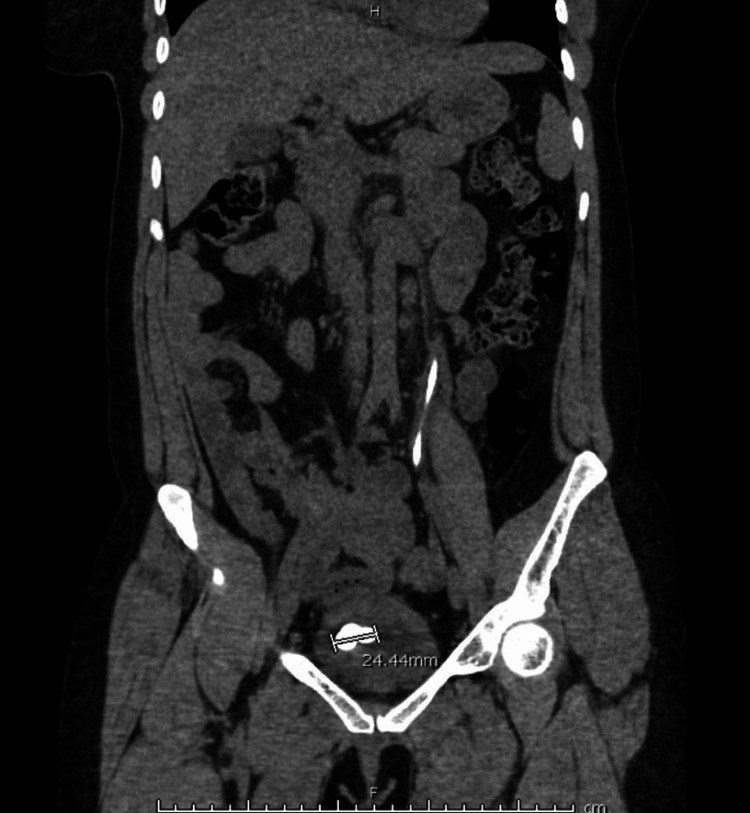
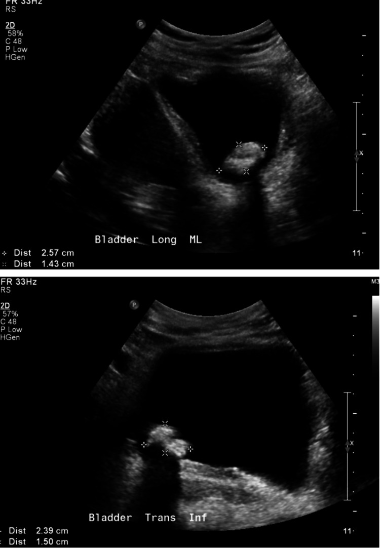
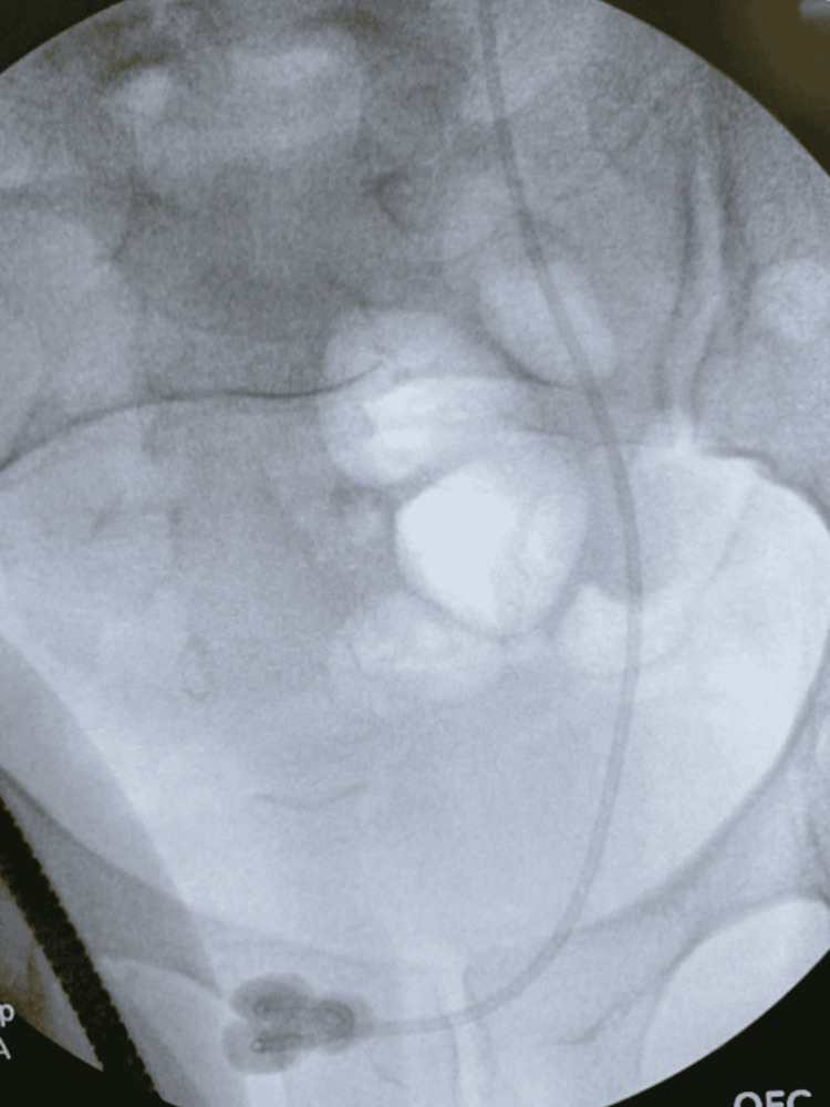
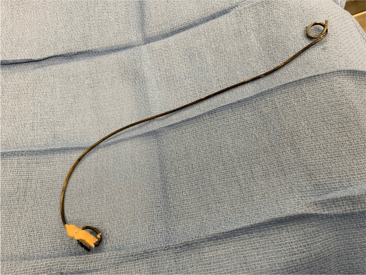
Double-J ureteral stents are an invaluable tool in urology and are one of the most widely used stents in the world. However, when left in situ for prolonged periods, so-called "retained" ureteral stents can lead to numerous complications such as migration, hematuria, encrustation, or stent occlusion. These complications present severe challenges in urologic management. Notably, encrustation of ureteral stents may increase the risk of renal impairment and other potentially life-threatening complications. Here, we present the case of a 34-year-old female with a left double-J ureteral stent who presented to the Emergency Department (ED) with a one-day history of left flank pain and febrile urinary tract infection.
Keywords: bladder calculi; endourology; lithotripsy; ureteral stent; urology.
Copyright © 2022, Aponte et al.
Conflict of interest statement
The authors have declared that no competing interests exist.
Figures
References
-
- Leslie SW, Sajjad H. StatPearls [Internet] Treasure Island (FL): StatPearls Publishing; 2022. Double J placement methods comparative analysis . - PubMed
-
- Ureteral stent encrustation: epidemiology, pathophysiology, management and current technology. Tomer N, Garden E, Small A, Palese M. https://doi.org/10.1097/ju.0000000000001343. J Urol. 2021;205:68–77. - PubMed
-
- Endourologic management of severely encrusted ureteral stents. Lojanapiwat B. http://www.jmatonline.com/index.php/jmat/article/view/12181#. J Med Assoc Thai. 2005;88:1203. - PubMed
-
- Endourological management of severely encrusted ureteral stents. Mohan-Pillai K, Keeley FX Jr, Moussa SA, Smith G, Tolley DA. J Endourol. 1999;13:377–379. - PubMed
Publication types
LinkOut - more resources
Full Text Sources