. 2022 Oct 12:9:935740.
doi: 10.3389/fnut.2022.935740. eCollection 2022.

Association study between hypothalamic functional connectivity, early nutrition, and glucose levels in healthy children aged 6 years: The COGNIS study follow-up

Affiliations

Association study between hypothalamic functional connectivity, early nutrition, and glucose levels in healthy children aged 6 years: The COGNIS study follow-up

Estefanía Diéguez et al. Front Nutr. .

Abstract

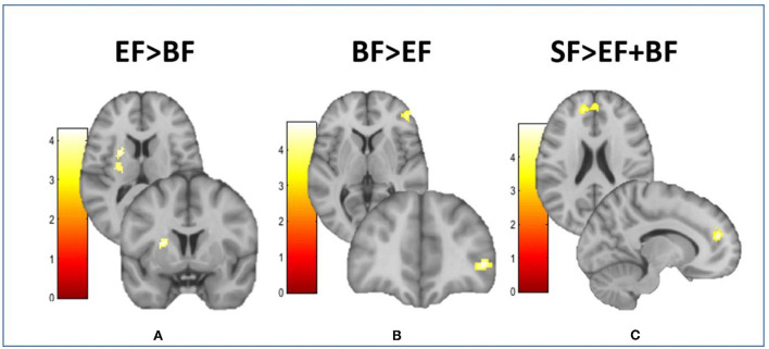
Breastfeeding (BF) is the gold standard in infant nutrition; knowing how it influences brain connectivity would help understand the mechanisms involved, which would help close the nutritional gap between infant formulas and breast milk. We analyzed potential long-term differences depending on the diet with an experimental infant formula (EF), compared to a standard infant formula (SF) or breastfeeding (BF) during the first 18 months of life on children's hypothalamic functional connectivity (FC) assessed at 6 years old. A total of 62 children participating in the COGNIS randomized clinical trial (Clinical Trial Registration: www.ClinicalTrials.gov, identifier: NCT02094547) were included in this study. They were randomized to receive an SF (n = 22) or a bioactive nutrient-enriched EF (n = 20). BF children were also included as a control study group (BF: n = 20). Brain function was evaluated using functional magnetic resonance imaging (fMRI) and mean glucose levels were collected through a 24-h continuous glucose monitoring (CGM) device at 6 years old. Furthermore, nutrient intake was also analyzed during the first 18 months of life and at 6 years old through 3-day dietary intake records. Groups fed with EF and BF showed lower FC between the medial hypothalamus (MH) and the anterior cingulate cortex (ACC) in comparison with SF-fed children. Moreover, the BF children group showed lower FC between the MH and the left putamen extending to the middle insula, and higher FC between the MH and the inferior frontal gyrus (IFG) compared to the EF-fed children group. These areas are key regions within the salience network, which is involved in processing salience stimuli, eating motivation, and hedonic-driven desire to consume food. Indeed, current higher connectivity found on the MH-IFG network in the BF group was associated with lower simple sugars acceptable macronutrient distribution ranges (AMDRs) at 6 months of age. Regarding linoleic acid intake at 12 months old, a negative association with this network (MH-IFG) only in the BF group was found. In addition, BF children showed lower mean glucose levels compared to SF-fed children at 6 years old. Our results may point out a possible relationship between diet during the first 18 months of life and inclined proclivity for hedonic eating later in life.

Clinical trial registration: https://www.clinicaltrials.gov/, identifier NCT02094547.

Keywords: eating behavior; hypothalamus; long chain polyunsaturated fatty acids (LC-PUFAs); mean glucose levels; milk fat globule membrane (MFGM); neuroimaging; synbiotics.


Conflict of interest statement

Authors JJ and RD-C are employees of Ordesa Laboratories, S.L. The remaining authors declare that the research was conducted in the absence of any commercial or financial relationships that could be construed as a potential conflict of interest.

Figures

Figure 1
Figure 1
Participant flowchart from baseline visit to 6 years old. BF, breastfeeding; D, dropouts; E, exclusions; EF, experimental infant formula; fMRI, functional magnetic resonance imaging; n, sample size; SF, standard infant formula. Up to 18 months of life, a total of 40 infants were excluded in the groups fed with SF and EF as follows: 24 were excluded in the SF-fed group (1 due to perinatal hypoxia, 1 had growth deficiency, not related to the infant formula, 15 did not take the infant formula, 2 had colic, 3 were excluded due to lactose intolerance, 1 was excluded due to digestive surgical intervention, and 1 suffered from hydrocephalus); 16 were excluded in the EF-fed group (2 presented growth deficiency, not related to the infant formula, 2 had lactose intolerance, 11 did not take the infant formula, and 1 was excluded due to epileptic seizure). While in the BF group, one infant was excluded, because he was not exclusively breastfed beyond the 2 months of age, when he started to take only infant formula. In the follow-up visits, drop-outs were due to participants' parents deciding to withdraw from the study. After drop-outs, *110 children (SF: 39; EF: 39; BF: 32) attended the follow-up visit at 6 years old. Nonetheless, not all parents attending to the follow-up visit wanted their children to participate in the fMRI session or wear the 24-h continuous glucose monitoring device. Moreover, according to methodology previously described (42), some fMRI data were eliminated because image quality was not adequate due to excessive movement inside the scanner (SF: 15; EF: 19; BF: 4). In addition, mean glucose data were collected with a continuous glucose monitoring device for an average of 7 days. Those glucose data registered for <3 days were not included in final analysis (SF: 5; EF: 8; BF: 4). Finally, 62 children were included at 6 years old in the current analysis (SF: 22; EF: 20; BF: 20).
Figure 2
Figure 2
Differences between groups in the resting-state functional connectivity of the medial hypothalamus. This figure shows different brain images of the medial hypothalamus. Color bars represent the connectivity intensity value or t-value. (A) EF > BF: MH-putamen extending to insula; (B) BF > EF: MH-IFG; (C) SF > EF + BF: MH-dorsal ACC. ACC, anterior cingulate cortex; BF, breastfeeding; EF, experimental infant formula; IFG, inferior frontal gyrus; MH, medial hypothalamus; SF, standard infant formula. Refer to Table 2 for indicated brain regions.
Figure 3
Figure 3
Significant differences in dietary intake in COGNIS-children up to 18 months of life. (A) 6 months old; (B) 12 months old; (C) 18 months old. AMDR, acceptable macronutrient distribution ranges; BF, breastfeeding; CHs, carbohydrates; DRI, dietary reference intake; EF, experimental infant formula; kcal, kilocalories; SF, standard infant formula. Simple sugars AMDR has not been determined. Values were classified as adequate or excess according to the maximal intake level, ≤ 25% of total daily energy intake (48). Values not sharing the same suffix (ab) were significantly different in the Bonferroni post-hoc test. **p < 0.01, ***p < 0.001.
Figure 4
Figure 4
Associations between resting-state functional connectivity at 6 years old and early nutrition at 6 months (A) and 12 months (B) of age in the BF group. AMDR, acceptable macronutrient distribution ranges; BF, breastfed; IFG, inferior frontal gyrus; MH, medial hypothalamus.

Similar articles

Cited by

References

    1. Black MM. Impact of nutrition on growth, brain, and cognition. Nestle Nutr Inst Workshop Ser. (2018) 89:185–95. 10.1159/000486502 - DOI - PubMed
    1. Gao W, Lin W, Grewen K, Gilmore JH. Functional connectivity of the infant human brain: plastic and modifiable. Neurosci Rev J Bring Neurobiol Neurol Psychiatry. (2017) 23:169–84. 10.1177/1073858416635986 - DOI - PMC - PubMed
    1. Deoni SCL. Neuroimaging of the developing brain and impact of nutrition. Nestle Nutr Inst Workshop Ser. (2018) 89:155–74. 10.1159/000486500 - DOI - PubMed
    1. Deoni S, Dean D, Joelson S, O'Regan J, Schneider N. Early nutrition influences developmental myelination and cognition in infants and young children. Neuroimage. (2018) 178:649–59. 10.1016/j.neuroimage.2017.12.056 - DOI - PMC - PubMed
    1. Georgieff MK, Ramel SE, Cusick SE. Nutritional influences on brain development. Acta Paediatr Oslo Nor. (2018) 107:1310–21. 10.1111/apa.14287 - DOI - PMC - PubMed

Associated data