. 2022 Oct 26;8(10):1383-1392.
doi: 10.1021/acscentsci.2c00125. Epub 2022 Aug 17.

Antibody Recognition of Different Staphylococcus aureus Wall Teichoic Acid Glycoforms

Affiliations

Antibody Recognition of Different Staphylococcus aureus Wall Teichoic Acid Glycoforms

Cristina Di Carluccio et al. ACS Cent Sci. .

Abstract

Wall teichoic acids (WTAs) are glycopolymers decorating the surface of Gram-positive bacteria and potential targets for antibody-mediated treatments against Staphylococcus aureus, including methicillin-resistant (MRSA) strains. Through a combination of glycan microarray, synthetic chemistry, crystallography, NMR, and computational studies, we unraveled the molecular and structural details of fully defined synthetic WTA fragments recognized by previously described monoclonal antibodies (mAbs 4461 and 4497). Our results unveiled the structural requirements for the discriminatory recognition of α- and β-GlcNAc-modified WTA glycoforms by the complementarity-determining regions (CDRs) of the heavy and light chains of the mAbs. Both mAbs interacted not only with the sugar moiety but also with the phosphate groups as well as residues in the ribitol phosphate (RboP) units of the WTA backbone, highlighting their significant role in ligand specificity. Using elongated WTA fragments, containing two sugar modifications, we also demonstrated that the internal carbohydrate moiety of α-GlcNAc-modified WTA is preferentially accommodated in the binding pocket of mAb 4461 with respect to the terminal moiety. Our results also explained the recently documented cross-reactivity of mAb 4497 for β-1,3/β-1,4-GlcNAc-modified WTA, revealing that the flexibility of the RboP backbone is crucial to allow positioning of both glycans in the antibody binding pocket.


Conflict of interest statement

The authors declare no competing financial interest.

Figures

Figure 1
Figure 1
S. aureus WTA is composed of repeating ribitol-5-phosphate (RboP) residues that can be decorated on the C-2 position with d-alanine esters and N-acetyl-d-glucosamine (GlcNAc) residues. The biosynthesis enzymes TarS, TarM, and TarP decorate the RboP backbone at C4 with β-GlcNAc, at C4 with α-GlcNAc, and at C3 with β-GlcNAc, respectively, to create different WTA glycoforms.
Figure 2
Figure 2
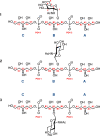
Ligands to probe antibody binding. The structures were designed to incorporate all structural features that were deemed important for mAb binding, a central glycosylated ribitol moiety flanked on each side by a RboP residue: (1) α-1,4-WTA; (2) β-1,4-WTA; (3) β-1,3-WTA.
Figure 3
Figure 3
(a) Schematic overview of the TA microarray library. Microarray results for (b) mAb 4497 and (c) mAb 4461 at different concentrations. Legend: 1 μg/mL, blue; 0.5 μg/mL, orange; 0.25 μg/mL, gray. Values refer to the average of the fluorescent median intensity of three spots for each compound printed at 30, 10, and 3 μM concentrations. n = number of repeating units.
Figure 4
Figure 4
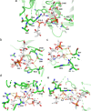
Different views of the binding site of IgG mAb 4461 in complex with (α-1,4)-GlcNAc WTA (ligand 1). (a) The α-GlcNAc-modified WTA 1 is bound in a cavity formed between the CDR of the heavy and light chains of the antibody. Residues S31, Y97, Y98, S100, and Y33 are involved in H-bond interactions with the ligand. (b) Residues Y97 and Y98 establish H-bonds with the GlcNAc ring, with the phosphate group 1, and with the Rbo-B3 OH. (c) Residue S100 forms a H-bond with the Rbo-A2 OH as well as the GlcNAc C4-OH and phosphate group 5 through a water molecule. Residue Y33 interacts via an H-bond with GlcNAc C6-OH. (d) Residues S31 and Y98 establish H-bonds with an oxygen atom of phosphate group 1. The ligand (α-1,4)-GlcNAc WTA 1 is shown in magenta.
Figure 5
Figure 5
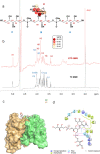
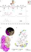
STD NMR and MD results of mAb 4461 and (α-1,4)-GlcNAc WTA, ligand 1 binding. (a) Epitope map of 1 interacting with mAb 4461 (only protons exhibiting %STD above 30% are indicated in the epitope mapping). (b) STD-NMR spectrum (red) of IgG 4461–1 and the unsaturated reference spectrum (black). (c) 3D view of the mAb4461–1 complex obtained from a molecular dynamics simulation. (d) Diagram of the interactions between mAb 4461 and 1 resulting from the best pose of the MD: solid arrows represent hydrogen bonds with functional groups of the amino acids of the antibody; the other residues in the binding pocket participate in polar and hydrophobic interactions.
Figure 6
Figure 6
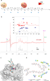
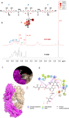
STD NMR and MD results of mAb 4461 and WTA hexamer 9 binding. (a) Epitope map of 9 interacting with IgG 4461. (b) STD-NMR spectrum (red) of mAb 4461–9 and the unsaturated reference spectrum (black). (c) 3D view of the mAb 4461–9 complex: the “internal” residue into the antibody binding site is shown in purple, and the “terminal” GlcNAc is shown in green. (d) Diagram of the interactions between mAb 4461 and 9 resulting from the manual docking: solid arrows represent hydrogen bonds with functional groups of the amino acids of the antibody; the other residues in the binding pocket participate in polar and hydrophobic interactions.
Figure 7
Figure 7
Different views of the binding site of IgG mAb 4497 in complex with ligand (β-1,4)-GlcNAc WTA ligand 2. (a) The β-GlcNAc-modified WTA is located in a cavity formed between the CDR of the heavy and light chains of the antibody. (b) Residues W33, D100, and G99 are involved in H-bond interactions with GlcNAc. Residue W33 stacks with the ligand and establishes a H-bond with the GlcNAc C4-OH. Residue G99 forms a H-bond with GlcNAc-C4-OH while D100 makes H-bonds with GlcNAc C3-OH and the Rbo-C4 hydroxyl group. (c) Residues N53 and S31 form H-bonds with GlcNAc C6-OH and phosphate group 1 via a water molecule. (d) Residue Y97 interacts with the GlcNAc C3-OH and the acetamide group while residue R31 interacts with phosphate group 5. (e) R34 establishes H-bonds with two oxygen atoms of phosphate group 5, with the Rbo-C4, Rbo-B3, and Rbo-A3 hydroxyl groups through two water molecules, and with the GlcNAc acetamide group. Finally, residue S33 forms a H-bond with Rbo-A3. Ligand 2 (β-1,4) is shown in white.
Figure 8
Figure 8
STD NMR and MD analysis of mAb 4497 and WTA-trimer 2 binding. (a) Epitope map of 2 interacting with IgG 4497 (only protons exhibiting %STD values above 30% are indicated in the epitope mapping). (b) STD-NMR spectrum (red) of mAb 4497–2 and the unsaturated reference spectrum (black). (c) 3D view of the mAb 4497–2 complex obtained from an MD simulation. (d) Description of the interactions between IgG 4497 and 2 resulting from a representative pose obtained by an MD simulation: solid arrows represent hydrogen bonds with functional groups of the amino acids of the antibody; the other residues in the binding pocket participate in polar and hydrophobic interactions.
Figure 9
Figure 9
Different views of the binding site of IgG mAb 4497 in complex with (β-1,3)-ligand 3. (a) The β-GlcNAc-modified WTA is located in a cavity formed between the CDR of the heavy and light chains of the antibody. (b) Residues D100, Y97, G99, S31, and W33 are involved in H-bond interactions with the GlcNAc. Residue W33 stacks with the GlcNAc and forms a H-bond with GlcNAc C4-OH. Residues G99, D100, and Y97 interact with the GlcNAc C4-OH and C3-OH, respectively. Residue D100 interacts with the Rbo-C4 hydroxyl group. (c) N53 is 4.1 Å from the GlcNAc C6-OH, while residue S31 interacts via H-bonds with GlcNAc C6-OH. Residue Y97 establishes a H-bond with the GlcNAc acetamide group and C3-OH. (d) R31 and R34 form H-bonds with both phosphate groups (1 and 5) and the acetamide group of GlcNAc. Finally, residue S33 establishes an H-bond with Rbo-A2. Ligand (β-1,3)-ligand 3 is shown in cyan.
Figure 10
Figure 10
Overlap of the ligands (β-1,4)-WTA trimer (2) and (β-1,3)-WTA trimer (3) within the binding site of IgG mAb 4497. (a) Overlap of ligands 2 and 3 within the binding site cavity of mAb IgG 4497. (b) Enlarged view of the overlap of the GlcNAc rings of ligands 2 and 3. (c) Lateral view of the overlapping ligands 1 and 2. Ligands 2 and 3 are shown in green and cyan respectively.
Figure 11
Figure 11
STD NMR and MD analysis of mAb 4497 and WTA-trimer 3 binding. (a) Epitope map of 3 interacting with mAb 4497 (only protons exhibiting %STD values above 30% are shown). (b) STD-NMR spectrum (red) of mAb 4497–3 and the unsaturated reference spectrum (blue). (c) 3D view of the mAb 4461–3 complex obtained from an MD simulation. (d) Description of the interactions between mAb 4497 and 3 resulting from a representative pose obtained by an MD simulation: solid arrows represent hydrogen bonds with functional groups of the amino acids of the antibody; the other residues in the binding pocket participate in polar and hydrophobic interactions.

References

    1. Krismer B.; Weidenmaier C.; Zipperer A.; Peschel A. The commensal lifestyle of Staphylococcus aureus and its interactions with the nasal microbiota.. Nat. Rev. Microbiol 2017, 15 (11), 675–687. 10.1038/nrmicro.2017.104. - DOI - PubMed
    1. Lehar S. M.; Pillow T.; Xu M.; Staben L.; Kajihara K. K.; Vandlen R.; DePalatis L.; Raab H.; Hazenbos W. L.; Hiroshi Morisaki J.; et al. S. Novel antibody-antibiotic conjugate eliminates intracellular S. aureus.. Nature 2015, 527 (7578), 323–328. 10.1038/nature16057. - DOI - PubMed
    1. Tacconelli E.; Carrara E.; Savoldi A.; Harbarth S.; Mendelson M.; Monnet D. L.; Pulcini C.; Kahlmeter G.; Kluytmans J.; Carmeli Y.; et al. Discovery, Research, and Development of New Antibiotics: The WHO Priority List of Antibiotic-Resistant Bacteria and Tuberculosis.. Lancet Infectious Diseases 2018, 18 (3), 318–327. 10.1016/S1473-3099(17)30753-3. - DOI - PubMed
    1. Weidenmaier C.; Peschel A. Teichoic acids and related cell-wall glycopolymers in Gram-positive physiology and host interactions.. A. Nat. Rev. Microbiol 2008, 6 (4), 276–287. 10.1038/nrmicro1861. - DOI - PubMed
    1. Kurokawa K.; Takahashi K.; Lee B. L. The staphylococcal surface-glycopolymer wall teichoic acid (WTA) is crucial for complement activation and immunological defense against Staphylococcus aureus infection.. Immunobiology 2016, 221 (10), 1091–1101. 10.1016/j.imbio.2016.06.003. - DOI - PubMed

LinkOut - more resources