. 2022 Oct 26;6(4):046101.
doi: 10.1063/5.0118311. eCollection 2022 Dec.

Long-acting acid-sensitive ketal-linked dexamethasone microcrystals for treating experimental autoimmune uveitis

Affiliations

Long-acting acid-sensitive ketal-linked dexamethasone microcrystals for treating experimental autoimmune uveitis

Maoyu Cai et al. APL Bioeng. .

Abstract

Corticosteroids have for some time been used as first-line drugs for the topical treatment of noninfectious uveitis, but poor ocular bioavailability and the rapid clearance of eye drops necessitate frequent dosing, reducing patient compliance. In this study, we used an acid-sensitive stearoxyl-ketal-dexamethasone pro-drug microcrystals (SKD MCs), which is consistently safe and effective in the control of uveitis inflammation in rats. We used a rat model of experimental autoimmune uveitis (EAU) to evaluate the effects of SKD MCs in terms of clinical manifestations, molecular biology, pathological histology, and visual electrophysiology compared to dexamethasone sodium phosphate injection or phosphate-buffered saline. SKD MCs significantly reduced inflammation in EAU, improved the ability to suppress inflammatory cytokines and to protect retinal function, and significantly reduced retinal microglia activation, with no increase in intraocular pressure throughout the treatment. Our results indicate that the SKD MCs formulation holds promise as a new strategy for the treatment of noninfectious uveitis and potentially other ocular inflammatory diseases.


Figures

FIG. 1.
FIG. 1.
Schematic illustration of the study protocol. Stearoxyl-ketal-dexamethasone pro-drug microcrystals (SKD MCs) were used to treat experimental autoimmune uveitis (EAU) in rats via a single subconjunctival injection. This study demonstrated that by suppressing the expression of inflammatory cytokines and activation of retinal microglia, SKD MCs achieved a long-lasting effect, with sustained reduction of inflammation. The right-hand panel shows three slit lamp images of anterior eyes (with dilated pupils) of rats with EAU on different treatments at day 18 post-immunization. In comparison to dexamethasone sodium phosphate (DSP) and phosphate-buffered saline (PBS), SKD MCs significantly reduced the dilatation of iris vessels and pupillary adhesions in EAU rats at day 18 post-immunization. In the central panel, red arrows indicate our processing of EAU rats (injection of SKD MCs into the subconjunctiva, and the results in EAU rats after different treatments). In the lower panel, yellow circles indicate stearyl alcohol, blue circles indicate dexamethasone, and distorted connecting lines indicate ketal bonds.
FIG. 2.
FIG. 2.
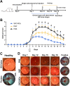
Therapeutic effect of stearoxyl-ketal-dexamethasone pro-drug microcrystal (SKD MCs) in an experimental anterior uveitis (EAU) rat model. (a) Schematic of EAU model establishment and treatment schedule. (b) Anterior segment disease score of rats in each group as a function of time. (c) Slit lamp anterior segment images and fundus images of healthy rats. (d) Slit lamp anterior segment images and fundus images of EAU rats under different treatments. Black stars indicate hypopyon, the black arrow indicates that the drug is continuously in place, and black triangles indicate tortuous dilatation of blood vessels in the fundus. *P <0.05 (PBS vs DSP); ∧P <0.05, (PBS vs SKD); #P <0.05 (SKD MCs vs DSP). Mean ± SEM, n = 8, one-way ANOVA test. PBS = phosphate-buffered saline; DSP = dexamethasone sodium phosphate.
FIG. 3.
FIG. 3.
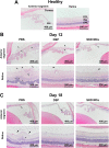
Effect of different treatments at different times on histopathologic changes in rats. Healthy rats without any intervention were used as controls (a). Histological images of the anterior segment and retina of each group of rats at day 12 (b) and day 18 (c) post-immunization, and corresponding histological images of healthy rats (a), with black arrows representing inflammatory cells and black stars representing distorted retinal structures. GCL = ganglion cell layer, INL = inner nuclear layer, ONL = outer nuclear layer.
FIG. 4.
FIG. 4.
Activation of retinal microglia under different treatments. (a) Effect of different treatment regimens on positive staining of retinal Iba1. The cell nuclei were stained using DAPI (blue) and the activated microglia in the retina were stained using Iba1 (green). (b) Retinal Iba1-positive cell density under different treatment regimens. *P <0.05, **P <0.01, ***P <0.001, and ****P <0.0001. Mean ± SEM, n = 4, one-way ANOVA test.
FIG. 5.
FIG. 5.
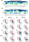
Protein and mRNA expression of inflammatory cytokines in different groups. Protein expression levels of 23 cytokines and chemokines detected by Luminex on day 12 post-immunization (a) and day 18 post-immunization (b). Expression of four inflammatory cytokine mRNAs in rat iris (c) and (d) and retinal (e) and (f) tissues on days 12 (c) and (e) and 18 (d) and (f) post-immunization. *P <0.05, **P <0.01, ***P <0.001, and ****P <0.0001. Mean ± SEM, n = 3, one-way ANOVA test.
FIG. 6.
FIG. 6.
Retinal function preservation in rats under different treatments. (a) Electroretinogram responses to stimuli at five intensity levels from four groups of rats. Relationship between flash stimulus intensity and a-wave mean amplitude (b) and b-wave mean amplitude (c). Asterisks represent significance of the difference at 2.5 log cd sec/m2. *P <0.05, **P <0.01, ***P <0.001, ****P <0.0001. Mean ± SEM, n = 4 eyes/group, two-way ANOVA test.

Similar articles

Cited by

References

    1. Miserocchi E. et al., “ Review on the worldwide epidemiology of uveitis,” Eur. J. Ophthalmol. 23, 705 (2013).10.5301/ejo.5000278 - DOI - PubMed
    1. de Smet M. D. et al., “ Understanding uveitis: The impact of research on visual outcomes,” Prog. Retinal Eye Res. 30, 452 (2011).10.1016/j.preteyeres.2011.06.005 - DOI - PubMed
    1. Suttorp-Schulten M. S. A. and Rothova A., “ The possible impact of uveitis in blindness: A literature survey,” Br. J. Ophthalmol. 80, 844 (1996).10.1136/bjo.80.9.844 - DOI - PMC - PubMed
    1. Pan J., Kapur M., and McCallum R., “ Noninfectious immune-mediated uveitis and ocular inflammation,” Curr. Allergy Asthma Rep. 14, 409 (2014).10.1007/s11882-013-0409-1 - DOI - PubMed
    1. Valdes L. M. and Sobrin L., “ Uveitis therapy: The corticosteroid options,” Drugs 80, 765 (2020).10.1007/s40265-020-01314-y - DOI - PubMed