. 2022 Oct 14:13:935418.
doi: 10.3389/fphar.2022.935418. eCollection 2022.

Neuroprotective effect of phospholipase A2 from Malaysian Naja sumatrana venom against H2O2-induced cell damage and apoptosis

Affiliations

Neuroprotective effect of phospholipase A2 from Malaysian Naja sumatrana venom against H2O2-induced cell damage and apoptosis

Nur Atiqah Haizum Abdullah et al. Front Pharmacol. .

Abstract

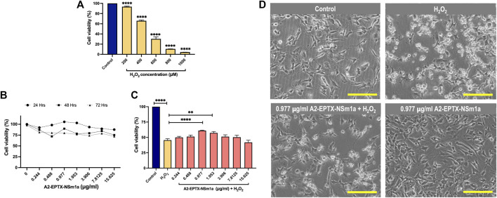
Oxidative stress is one of the factors involved in the pathogenesis of several neurodegenerative diseases. It has been reported that a secretory phospholipase A2 known as A2-EPTX-NSm1a has lower cytotoxicity in neuronal cells compared to its crude Naja sumatrana venom. In this study, A2-EPTX-NSm1a was tested for its neuroprotective activity on human neuroblastoma cells (SH-SY5Y) differentiated into cholinergic neurons against oxidative stress induced by hydrogen peroxide (H2O2). H2O2 treatment alone increased the caspase-3 and caspase-8 activities, whereas pre-treatment with A2-EPTX-NSm1a reduced the activity of these apoptosis-associated proteins. Moreover, A2-EPTX-NSm1a protects the morphology and ultrastructure of differentiated SH-SY5Y cells in the presence of H2O2. Oxidative stress increased the number of small mitochondria. Further evaluation showed the size of mitochondria with a length below 0.25 µm in oxidative stress conditions is higher than the control group, suggesting mitochondria fragmentation. Pre-treatment with A2-EPTX-NSm1a attenuated the number of mitochondria in cells with H2O2 Furthermore, A2-EPTX-NSm1a altered the expression of several neuroprotein biomarkers of GDNF, IL-8, MCP-1, TIMP-1, and TNF-R1 in cells under oxidative stress induced by H2O2. These findings indicate that anti-apoptosis with mitochondria-related protection, anti-inflammatory effect, and promote expression of important markers for cell survival may underlie the neuroprotective effect of A2-EPTX-NSm1a in cholinergic rich human cells under oxidative stress, a vital role in the neuronal disorder.

Keywords: apoptosis; inflammation; mitochondria; neurodegenerative disease; neuroprotection; snake venom phospholipase A2.


Conflict of interest statement

The authors declare that the research was conducted in the absence of any commercial or financial relationships that could be construed as a potential conflict of interest.

Figures

FIGURE 1
FIGURE 1
Differentiation of SH-SY5Y cells. (A) SH-SY5Y were differentiated as in the line chart. (B) Phase contrast of SH-SY5Y cells morphology on days 0, 4 and 8. Neurite extension labelled in red arrow. Scale bar = 200 μM. (C) Differentiation of SH-SY5Y showed modification in the cell number, (D) neurite length and (E) AChE activity. Data were presented as means ± SD, where p < 0.05 is considered significant. **** indicated p < 0.001.
FIGURE 2
FIGURE 2
Cell proliferation and neuroprotection of a secretory phospholipase A2, A2-EPTX-NSm1a in differentiated SH-SY5Y cell lines. (A) Cytotoxicity of H2O2 on differentiated SH-SY5Y cells. (B) Cell proliferation of A2-EPTXNSm1a was evaluated at different times of incubation. (C) Neuroprotection assay of A2-EPTX-NSm1a. (D) Phase contrast of differentiated SH-SY5Y in the different groups - untreated (control), H2O2 only (oxidative group), pre-treatment of 0.977 µg/ml A2-EPTX-NSm1a followed by H2O2 and 0.977 µg/ml A2-EPTX-NSm1a only after 24 h of incubation time. Scale bar = 200 μM.
FIGURE 3
FIGURE 3
Effect of pre-challenged A2-EPTX-NSm1a on caspase-3 and caspase-8 activity in differentiated SH-SY5Y with H2O2. (A) Relative Caspase-3 activity (fold change); (B) Relative Caspase-8 activity (fold change) All experiment was conducted in triplicate (n = 3). Data are presented as the mean ± SD. * p < 0.05, ** p < 0.01, *** p < 0.005, **** p < 0.001 as compared to the H2O2-treated group.
FIGURE 4
FIGURE 4
Representative cell structure images of differentiated SH-SY5Y observed in an electron microscope. Image of differentiated SH-SY5Y cells in (A) untreated group (Control), (B) H2O2-induced neurotoxicity group, (C) addition of pre-treatment of A2-EPTX-NSm1a following the addition of H2O2 and (D) A2-EPTX-NSm1a alone at 24 h of incubation. Scale bar = 1 μm. Arrow showed the formation of membrane bubbling due to cell apoptosis induced by H2O2.
FIGURE 5
FIGURE 5
Observation of mitochondrial morphology from electron microscope. (A) Morphology of mitochondria in each group observed after 24 h of treatment in a representative of cell. (B) Number of mitochondria in the area has been quantified and the length of mitochondria in each group were measured using ImageJ analysis. Scale bar = 200 nm. Data showed mean ± SEM and marked with ** indicating p < 0.01, *** as p < 0.005, and **** marked as p < 0.001.
FIGURE 6
FIGURE 6
A2-EPTX-NSma1 effects on the neuro-markers expression levels in the differentiated SH-SY5Y with and without oxidative stress conditions. The expression level of proteins was analysed using normalized signals extracted from Agilent Feature Extraction software. Values are collected from quadruplicates of a specific protein and expressed as mean ± standard deviation (n = 3). Protein expression is considered as significant as compared to the H2O2 group with p < 0.05 based on statistical analysis using one-way ANOVA followed by Tukey’s post-test. ns as significant, * p < 0.05, ** p < 0.01, *** p < 0.005, **** p < 0.0001.

References

    1. Aarsland D., Creese B., Politis M., Chaudhuri K. R., Ffytche D. H., Weintraub D., et al. (2017). Cognitive decline in Parkinson disease. Nat. Rev. Neurol. 13 (4), 217–231. 10.1038/nrneurol.2017.27 - DOI - PMC - PubMed
    1. Abdullah N. A. H., Rusmili M. R. A., Zainal Abidin S. A., Shaikh M. F., Hodgson W. C., Othman I. (2021). Isolation and characterization of A2-EPTX-nsm1a, a secretory phospholipase A2 from Malaysian spitting cobra (Naja sumatrana) venom. Toxins 13 (12), 859. 10.3390/toxins13120859 - DOI - PMC - PubMed
    1. Akhter N., Wilson A., Thomas R., Al-Rashed F., Kochumon S., Al-Roub A., et al. (2021). ROS/TNF-α crosstalk triggers the expression of IL-8 and MCP-1 in human monocytic THP-1 cells via the NF-κB and ERK1/2 mediated signaling. Int. J. Mol. Sci. 22 (19), 10519. 10.3390/ijms221910519 - DOI - PMC - PubMed
    1. Armugam A., Cher C. D., Lim K., Koh D. C., Howells D. W., Jeyaseelan K. (2009). A secretory phospholipase A2-mediated neuroprotection and anti-apoptosis. BMC Neurosci. 10 (120), 120–213. 10.1186/1471-2202-10-12010.1186/1471-2202-10-120 - DOI - PMC - PubMed
    1. Bachiller S., Jiménez-Ferrer I., Paulus A., Yang Y., Swanberg M., Deierborg T., et al. (2018). Microglia in neurological diseases: A road map to brain-disease dependent-inflammatory response. Front. Cell. Neurosci. 12, 488. 10.3389/fncel.2018.00488 - DOI - PMC - PubMed