

Predictive value of myocardial strain on myocardial infarction size by cardiac magnetic resonance imaging in ST-segment elevation myocardial infarction with preserved left ventricular ejection fraction
- PMID: 36313364
- PMCID: PMC9613930
- DOI: 10.3389/fphar.2022.1015390
Predictive value of myocardial strain on myocardial infarction size by cardiac magnetic resonance imaging in ST-segment elevation myocardial infarction with preserved left ventricular ejection fraction
Abstract
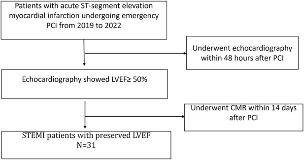
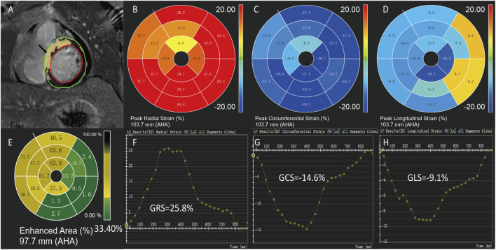
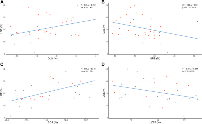
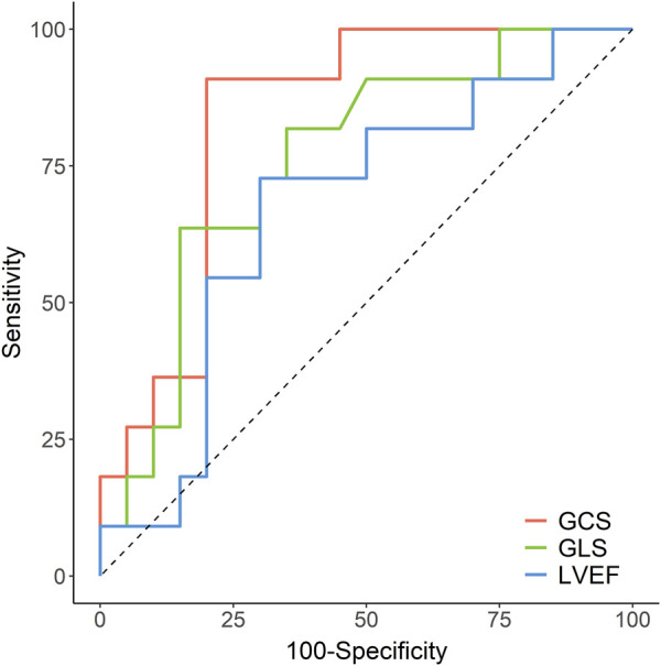
Background: The correlation between myocardial strain and infraction size by cardiac magnetic resonance imaging in ST-segment elevation myocardial infarction (STEMI) with preserved left ventricular ejection fraction (LVEF) is not clear. Objective: To investigate the correlation between myocardial strain and myocardial infarction size in patients of acute STEMI with preserved LVEF. Materials and Methods: A retrospective study was conducted to assess 31 patients with acute ST-segment elevation myocardial infarction (STEMI)after primary percutaneous coronary intervention (PCI) who received cardiac magnetic resonance (CMR) imaging during hospitalization at the Central Hospital of Shandong First Medical University from 2019 to 2022 and whose echocardiography indicated preserved LVEF (LVEF≥50%). The control group consisted of 21 healthy adults who underwent CMR during the same period. We compared the CMR characteristics, global and segmental strain between the two groups. Furthermore, the correlation between the global strain and the segmental strain of the left ventricle and late gadolinium enhancement (LGE) were evaluated. Results: Compared with healthy controls, the left ventricular ejection fraction (LVEF) of STEMI patients with preserved LVEF was significantly decreased (p < 0.05). Moreover, the global radial strain (GRS) (24.09% [IQR:17.88-29.60%] vs. 39.56% [IQR:29.19-42.20%], p < 0.05), global circumferential strain (GCS) [-14.66% (IQR: 17.91-11.56%) vs. -19.26% (IQR: 21.03-17.73%), p < 0.05], and global longitudinal strains (GLS) (-8.88 ± 2.25% vs. -13.46 ± 2.63%, p < 0.05) were damaged in patients. Furthermore, GCS and GLS were associated with LGE size (%left ventricle) (GCS: r = 0.58, p < 0.05; GLS: r = 0.37, p < 0.05). In the multivariate model, we found that LGE size was significantly associated with GCS (β coefficient = 2.110, p = 0.016) but was not associated with GLS (β coefficient = -0.102, p = 0.900) and LVEF (β coefficient = 0.227, p = 0.354). The receiver operating characteristic (ROC) results showed that GCS emerged as the strongest LGE size (LGE >25%) prognosticator among strain parameters (AUC: 0.836 [95% CI, 0.692-0.981], sensitivity: 91%, specificity: 80%) and was significantly better (p = 0.001) than GLS [AUC: 0.761 (95% CI, 0.583-0.939), sensitivity: 64%, specificity: 85%] and LVEF [AUC: 0.673 (95% CI, 0.469-0.877), sensitivity: 73%, specificity: 70%]. Conclusion: Among STEMI patients with preserved LVEF after PCI, CMR-FT-derived GCS had superior diagnostic accuracy than GLS and LVEF in predicting myocardial infarction size.
Keywords: acute ST-segment elevation myocardial infarction; late gadolinium enhancement (LGE); magnetic resonance imaging; preserved left ventricle ejection fraction; strain.
Copyright © 2022 Wang, Wang, Ma, Wang, Li, Tian, Yue, Su and Li.
Conflict of interest statement
Author XY was employed by the company Philips Healthcare. The remaining authors declare that the research was conducted in the absence of any commercial or financial relationships that could be construed as a potential conflict of interest.
Figures
Similar articles
-
[Predictive value of global longitudinal strain measured by cardiac magnetic resonance imaging for left ventricular remodeling after acute ST-segment elevation myocardial infarction: a multi-centered prospective study].Nan Fang Yi Ke Da Xue Xue Bao. 2024 Jun 20;44(6):1033-1039. doi: 10.12122/j.issn.1673-4254.2024.06.03. Nan Fang Yi Ke Da Xue Xue Bao. 2024. PMID: 38977332 Free PMC article. Chinese.
-
Novel insights into myocardial fibrosis in patients with new onset ST-elevation myocardial infarction following percutaneous coronary intervention through enhanced cardiac magnetic resonance imaging: a prospective cohort study.BMC Cardiovasc Disord. 2025 Apr 10;25(1):274. doi: 10.1186/s12872-025-04719-3. BMC Cardiovasc Disord. 2025. PMID: 40211110 Free PMC article.
-
Left ventricular flow kinetics and myocardial deformation following acute infarction: Additional predictive value of cardiac magnetic resonance four-dimensional flow for left ventricular remodeling post-ST-elevation myocardial infarction.J Cardiovasc Magn Reson. 2025 May 7;27(2):101905. doi: 10.1016/j.jocmr.2025.101905. Online ahead of print. J Cardiovasc Magn Reson. 2025. PMID: 40345668 Free PMC article.
-
Assessing Myocardial Strain and Myocardial Work as a Marker for Hypertensive Heart Disease: A Meta-Analysis.Rev Cardiovasc Med. 2023 Jul 31;24(8):217. doi: 10.31083/j.rcm2408217. eCollection 2023 Aug. Rev Cardiovasc Med. 2023. PMID: 39076705 Free PMC article.
-
Prognostic impact of MRI-derived feature tracking myocardial strain in patients with non-ischaemic dilated cardiomyopathy: a systematic review and meta-analysis.Clin Radiol. 2024 May;79(5):e702-e714. doi: 10.1016/j.crad.2023.12.029. Epub 2024 Feb 13. Clin Radiol. 2024. PMID: 38402086
Cited by
-
Association between cardiac magnetic resonance ventricular strain and left ventricular thrombus in patients with ST-segment elevation myocardial infarction.Int J Cardiovasc Imaging. 2024 Aug;40(8):1735-1744. doi: 10.1007/s10554-024-03163-2. Epub 2024 Jun 17. Int J Cardiovasc Imaging. 2024. PMID: 38884697
-
Right ventricular function and determining factors of dysfunction in ST-segment-elevation myocardial infarction: a cross-sectional study with cardiac magnetic resonance imaging (MRI).Quant Imaging Med Surg. 2024 Sep 1;14(9):6895-6907. doi: 10.21037/qims-23-1804. Epub 2024 Jun 11. Quant Imaging Med Surg. 2024. PMID: 39281121 Free PMC article.
References
-
- Bodi V., Sanchis J., Nunez J., Mainar L., Lopez-Lereu M. P., Monmeneu J. V., et al. (2009). Prognostic value of a comprehensive cardiac magnetic resonance assessment soon after a first ST-segment elevation myocardial infarction. JACC. Cardiovasc. Imaging 2 (7), 835–842. 10.1016/j.jcmg.2009.03.011 - DOI - PubMed
-
- Bogaert J., Bosmans H., Maes A., Suetens P., Marchal G., Rademakers F. E. (2000). Remote myocardial dysfunction after acute anterior myocardial infarction: Impact of left ventricular shape on regional function: A magnetic resonance myocardial tagging study. J. Am. Coll. Cardiol. 35 (6), 1525–1534. 10.1016/s0735-1097(00)00601-x - DOI - PubMed
-
- Cerqueira M. D., Weissman N. J., Dilsizian V., Jacobs A. K., Kaul S., Laskey W. K., et al. (2002). Standardized myocardial segmentation and nomenclature for tomographic imaging of the heart. A statement for healthcare professionals from the Cardiac Imaging Committee of the Council on Clinical Cardiology of the American Heart Association. J. Nucl. Cardiol. 9 (2), 240–245. 10.1067/mnc.2002.123122 - DOI - PubMed
LinkOut - more resources
Full Text Sources
Miscellaneous