. 2022 Oct 13:10:1018161.
doi: 10.3389/fcell.2022.1018161. eCollection 2022.

Increased Aurora B expression reduces substrate phosphorylation and induces chromosomal instability

Affiliations

Increased Aurora B expression reduces substrate phosphorylation and induces chromosomal instability

Eric M C Britigan et al. Front Cell Dev Biol. .

Abstract

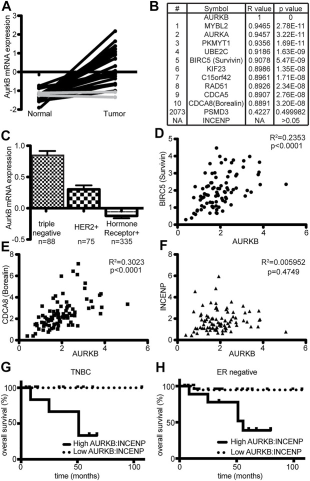
Increased Aurora B protein expression, which is common in cancers, is expected to increase Aurora B kinase activity, yielding elevated phosphorylation of Aurora B substrates. In contrast, here we show that elevated expression of Aurora B reduces phosphorylation of six different Aurora B substrates across three species and causes defects consistent with Aurora B inhibition. Complexes of Aurora B and its binding partner INCENP autophosphorylate in trans to achieve full Aurora B activation. Increased expression of Aurora B mislocalizes INCENP, reducing the local concentration of Aurora B:INCENP complexes at the inner centromere/kinetochore. Co-expression of INCENP rescues Aurora B kinase activity and mitotic defects caused by elevated Aurora B. However, INCENP expression is not elevated in concert with Aurora B in breast cancer, and increased expression of Aurora B causes resistance rather than hypersensitivity to Aurora B inhibitors. Thus, increased Aurora B expression reduces, rather than increases, Aurora B kinase activity.

Keywords: CIN; aurora kinase inhibitor; mitosis; mitotic checkpoint; spindle assembly checkpoint.


Conflict of interest statement

M.E.B is a member of the Medical advisory board of Strata Oncology and receives Research funding from Abbvie, Genentech, Puma, Arcus, Apollomics, Loxo Oncology/Lilly, Seagen and Elevation Oncology. The remaining authors declare that the research was conducted in the absence of any commercial or financial relationships that could be construed as a potential conflict of interest.

Figures

FIGURE 1
FIGURE 1
ARF loss results in increased Aurora B protein but reduced Aurora B activity. (A) Immunoblot of ARF+/+ and ARF−/− MEFs showing Aurora B is increased in ARF−/− MEFs while pT237 (active) Aurora B is decreased. Coomassie is shown as a loading control. Quantitation is shown below. (B–E) Quantitative immunofluorescence of pH3 and pDSN1 in ARF+/+ and ARF−/− MEFs. (B,D) Representative images of pH3 (B) and pDSN1 (D) in ARF+/+ and ARF−/− MEFs. (C,E) Graphs showing quantification of pH3 (C) and pDSN1 (D). n > 10 cells from each of 3 independent experiments. *p < 0.05.
FIGURE 2
FIGURE 2
Expression of Aurora B-YFP reduces Aurora B-dependent phosphorylation events in primary MEFs. (A) Immunoblot of MEFs of the indicated genotypes±Aurora B-YFP. ARF−/− MEFs show increased Aurora B and decreased pT237 (active) Aurora B. Coomassie (bottom) shown as loading control. (B–E) Quantitative immunofluorescence of pH3 and pDSN1. (B,D) Representative images of ARF+/+ MEFs transfected with YFP or Aurora B-YFP. H2B-YFP was co-transfected and used as a marker of transfected cells. (C,E) Graphs showing quantification of phosphoantibody signal in transfected cells. n > 10 cells from each of 3 independent experiments. *p < 0.05.
FIGURE 3
FIGURE 3
Expression of GFP-Aurora B in HeLa cells causes a reduction in Aurora B kinase activity and an increase in mitotic defects. Cells were analyzed 48 h after infection with retroviruses expressing GFP-Aurora B or GFP alone. (A) Immunoblot showing endogenous pT232 (active) Aurora B is reduced in HeLa cells overexpressing Aurora B. Tubulin is shown as a loading control. (B) Immunoblot showing reduced phosphorylation of the bona fide Aurora B substrate histone H3 Serine 10 in HeLa cells infected with retroviruses expressing GFP-Aurora. Tubulin is shown as a loading control. (C–H) Quantitative immunofluorescence demonstrating that expression of GFP-Aurora B decreases phosphorylation of histone H3 (pH3), the kinetochore component DSN1, and INCENP in HeLa cells. (C,E,G) Representative images of pH3, pDSN1, and pINCENP, respectively, in HeLa cells expressing GFP and GFP-Aurora B. (D,F,H) Quantification of pH3 S10 (D), pDSN1 S109 (F), and pINCENP TSS (H) demonstrating that expression of GFP-Aurora B significantly reduces phosphorylation of these Aurora B substrates in HeLa cells. n > 10 cells from each of 3 independent experiments. (I–P) GFP-Aurora B expression increases the incidence of mitotic defects. (I) Representative image of a misaligned chromosome (arrow). (J) Expression of GFP-Aurora B in HeLa cells causes an increase in misaligned chromosomes in metaphase. n > 50 metaphase cells from 3 independent experiments. (K) Representative image of a lagging chromosome (arrow). (L) Expression GFP-Aurora B in HeLa cells causes an increase in lagging chromosomes. n > 250 anaphase and telophase cells from each of 3 independent experiments. (M) Representative image of a multipolar spindle. (N) Expression of GFP-Aurora B in HeLa cells causes an increase in multipolar spindles. n > 250 mitotic cells from each of 3 independent experiments. (O) Expression of GFP-Aurora B increases the percentage of binucleate HeLa cells. n > 250 cells from each of three independent experiments. (P) Expression of GFP-Aurora B in HeLa cells impairs their ability to arrest in mitosis after 16 h of treatment with the microtubule poison colcemid, consistent with a weakened mitotic checkpoint. n > 250 cells from each of 3 independent experiments. *p < 0.05.
FIGURE 4
FIGURE 4
Increased expression of untagged Aurora B inhibits Aurora B kinase activity and causes resistance to Aurora kinase inhibition. (A) Immunoblot showing ≥2- fold overexpression of untagged Aurora B in MDA-MB-231 cells treated with tetracycline. Despite increased protein levels of Aurora B, pT232 (active) Aurora B is reduced. Tubulin is shown as a loading control. (B–E) Quantitative immunofluorescence showing that increased Aurora B expression results in reduced phosphorylation of the Aurora B substrates histone H3 and KNL1. (B,D) Representative images of pH3 and pKNL1 in MDA-MB-231 cells ± 48 h of tet-inducible expression of Aurora B (C,E) Quantification of B and D. n > 10 cells from each of 3 independent experiments. (F–I) 48 h of tet-induced Aurora B expression induces mitotic defects in MDA-MB-231 cells. (F) Representative image of a misaligned chromosome (arrow). (G) Elevated Aurora B expression increases the incidence of misaligned chromosomes. n > 50 metaphases from 3 independent experiments. (H) Representative image of a lagging chromosome (arrow). (I) Increased Aurora B expression induces lagging chromosomes. n > 100 anaphase and telophases from 3 independent experiments. (J–O) Increased Aurora B expression causes resistance to Aurora B inhibition. (J) Primary ARF−/− MEFs grow significantly better than ARF+/+ MEFS after 16 h of exposure to the Aurora B inhibitor ZM447439. Cell number shown is normalized to the number of ARF+/+ cells 10 days after treatment with DMSO. n = 3 independent experiments. (K) Primary ARF−/− MEFs (red), which overexpress endogenous Aurora B, have significantly improved colony forming ability as compared to ARF+/+ MEFs (blue) after 16 h exposure to ZM447439. n = 3 independent experiments. (L) MDA-MB-231 cells expressing untagged Aurora B in response to tetracycline grow significantly better over the course of 10 days than controls after 16 h exposure to ZM447439. n = 3 independent experiments. (M) MDA-MB-231 cells expressing tet-inducible wild type Aurora B exhibit significantly elevated colony forming ability compared to control MDA-MB-231 cells after exposure to ZM447439. n = 3 independent experiments. (N) MCF7 cells expressing Aurora B–GFP in response to tetracycline grow significantly better over the course of 10 days than MCF7 controls after treatment with ZM447439. n = 3 independent experiments. (O) MCF7 cells expressing Aurora B–GFP form significantly more colonies than control cells after exposure to ZM447439. n = 3 independent experiments. *p < 0.05.
FIGURE 5
FIGURE 5
Co-expression of INCENP restores Aurora B kinase activity and rescues mitotic defects. (A–D) Upregulation of Aurora B mislocalizes INCENP. (A) ARF−/− MEFs, which have increased levels of endogenous Aurora B, have reduced INCENP localization to the inner centromere. (B) Quantification of INCENP levels in primary MEFs. n > 10 cells from each of 3 independent experiments. (C) Tet-inducible Aurora B expression in MDA-MB-231 cells reduces INCENP localization to the inner centromere. (D) Quantification of INCENP levels in MDA-MB-231 cells. n > 10 cells from each of 3 independent experiments. (E–L) Infection with lentiviruses expressing INCENP-mNeonGreen (48 h) rescues Aurora B inhibition and mitotic defects caused by tet-induced upregulation of Aurora B (48 h). (E–H) Aurora B upregulation reduces phosphorylation of Aurora B substrates, but co-expression of INCENP-mNeonGreen restores phosphorylation of pH3 (E–F) and pKNL1 (G–H). (F,H) Quantification of pH3 (F) and pKNL (H) levels. n > 10 cells from each of 3 independent experiments. (I–J) Upregulation of Aurora B increases the incidence of misaligned (I) and lagging (J) chromosomes, while expression of INCENP-mNeonGreen in conjunction with Aurora B upregulation rescues this defect. n > 50 metaphase cells (I) or 100 anaphase and telophase cells (J) from 3 independent experiments. (K) Aurora B overexpression increases the incidence of multipolar spindles, but co-expression of INCENP rescues this defect. n > 250 mitotic cells from each of 3 independent experiments. (L) Upregulation of Aurora B impairs mitotic checkpoint signaling, resulting in a reduced mitotic index after 16 h of colcemid treatment. Expression of INCENP-mNeonGreen in conjunction with Aurora B upregulation rescues this defect. n > 250 cells from each of 3 independent experiments. (M) ARF−/− MEFs, which have elevated protein levels of endogenous Aurora B, have an impaired ability to arrest in mitosis after 16 h of treatment with the microtubule poison colcemid. However, expression of INCENP-mNeonGreen restores mitotic checkpoint signaling in ARF−/− MEFs. n > 250 cells from each of 3 independent experiments. *p < 0.05.
FIGURE 6
FIGURE 6
Expression of INCENP is sufficient to increase Aurora B activity in cells endogenously expressing elevated levels of Aurora B. (A–C) Schematic diagram of CRISPR-Cas9 p14ARF knockout (KO) strategy. (A) The CDKN2A genomic locus encodes for p14ARF and p16INKA. Two guide RNAs targeting exon1β of CDKN2A were used to produce specific p14ARF KO. (B) PCR strategy for confirming successful KO of p14ARF. (C) MDA-MB-231 cells expressing Cas9-P2A-NeonGreen were transiently transfected with a construct expressing mScarlet and multiple guide RNAs and FACs sorted for mScarlet-NeonGreen double positive cells before genomic analysis for p14ARF knockout. (D) Double positive cells were screened using the PCR strategy in (B). Homozygous KO clones show a 600 bp DNA band. (E) p14ARF KO clones 2 and 12 show complete KO of p14ARF by immunoblot, which induces constitutively elevated levels of endogenous Aurora B and lower levels of pH3. (F) p14ARF KO increases Aurora B and reduces phosphorylation of Aurora B and pH3. Expression of INCENP-mScarlet restores endogenous Aurora B kinase activity.
FIGURE 7
FIGURE 7
Aurora B expression correlates with Survivin and Borealin, but not INCENP, in breast cancer. Analysis of TCGA breast cancer data (Network, 2012). (A) Analysis of AURKB (Aurora B) mRNA expression levels in 22 matched pairs of normal and cancerous breast tissue. Lines connect dots displaying the AURKB expression level in matching tissue. In 20 of 22 cases, expression of AURKB mRNA is substantially higher in tumor tissue than in matched normal breast tissue (black). The other two cases are shown in gray. (B) Pavlidis Template Matching was used to identify genes whose expression most strongly correlates with AURKB in the 22 tumor samples in (A). Expression of BIRC5 (Survivin) and CDCA8 (Borealin) is highly correlated with that of AURKB. INCENP is absent from the 468 genes whose expression correlated with AURKB to a corrected p value < 0.05. The full list of genes is shown in Supplementary Table S1. (C) Stratification of all breast cancers classified with respect to subtype showing that AURKB is most highly expressed in triple negative breast cancers. (D–F) Dot plots of mRNA expression values of AURKB with BIRC5 (D), CDCA8 (E), and INCENP (F) showing that AURKB expression correlates with BIRC5 and CDCA8 but not with INCENP in triple negative breast cancers. Pavlidis Template Matching further demonstrates that BIRC5 and CDCA8 expression correlate with AURKB, while INCENP expression does not (see Supplementary Table S2). (G) Kaplan-Meier curves for overall patient survival of high or low AURKB:INCENP ratio expressing tumors. Triple Negative Breast Cancers (TNBC) were divided into those exhibiting an AURKB:INCENP protein ratio of >1.2 or less than 1.2. TNBC tumors with an AURKB:INCENP ratio >1.2 have significantly more aggressive disease that those with a ratio <1.2 (Log Rank test p = 0.0376). (H) Same as in (G) but using histologically ER-tumors. ER-tumors with a high AURKB:INCENP ratio show an aggressive disease progression (p = 0.0253).

Similar articles

Cited by

References

    1. Adams R. R., Wheatleya S. P., Gouldsworthy A. M., Kandels-Lewis S. E., Carmena M., Smythe C., et al. (2000). INCENP binds the Aurora-related kinase AIRK2 and is required to target it to chromosomes, the central spindle and cleavage furrow. Curr. Biol. 10 (17), 1075–1078. 10.1016/s0960-9822(00)00673-4 - DOI - PubMed
    1. Adams R. R., Maiato H., Earnshaw W. C., Carmena M. (2001). Essential roles of Drosophila inner centromere protein (INCENP) and aurora B in histone H3 phosphorylation, metaphase chromosome alignment, kinetochore disjunction, and chromosome segregation. J. Cell. Biol. 153 (4), 865–880. 10.1083/jcb.153.4.865 - DOI - PMC - PubMed
    1. Altieri D. C. (2008). New wirings in the survivin networks. Oncogene 27 (48), 6276–6284. 10.1038/onc.2008.303 - DOI - PMC - PubMed
    1. Azzariti A., Bocci G., Porcelli L., Fioravanti A., Sini P., Simone G. M., et al. (2011). Aurora B kinase inhibitor AZD1152: Determinants of action and ability to enhance chemotherapeutics effectiveness in pancreatic and colon cancer. Br. J. Cancer 104 (5), 769–780. 10.1038/bjc.2011.21 - DOI - PMC - PubMed
    1. Biggins S., Murray A. W. (2001). The budding yeast protein kinase Ipl1/Aurora allows the absence of tension to activate the spindle checkpoint. Genes. Dev. 15 (23), 3118–3129. 10.1101/gad.934801 - DOI - PMC - PubMed

LinkOut - more resources