. 2022 Oct 14:16:948327.
doi: 10.3389/fncel.2022.948327. eCollection 2022.

Artificial sleep-like up/down-states induce synaptic plasticity in cortical neurons from mouse brain slices

Affiliations

Artificial sleep-like up/down-states induce synaptic plasticity in cortical neurons from mouse brain slices

Gai-Linn Kay Besing et al. Front Cell Neurosci. .

Abstract

During non-rapid eye movement (NREM) sleep, cortical neuron activity alternates between a depolarized (firing, up-state) and a hyperpolarized state (down-state) coinciding with delta electroencephalogram (EEG) slow-wave oscillation (SWO, 0. 5-4 Hz) in vivo. Recently, we have found that artificial sleep-like up/down-states can potentiate synaptic strength in layer V cortical neurons ex vivo. Using mouse coronal brain slices, whole cell voltage-clamp recordings were made from layer V cortical pyramidal neurons to record spontaneous excitatory synaptic currents (sEPSCs) and inhibitory synaptic currents (sIPSCs). Artificial sleep-like up/down-states (as SWOs, 0.5 Hz, 10 min, current clamp mode) were induced by injecting sinusoidal currents into layer V cortical neurons. Baseline pre-SWO recordings were recorded for 5 min and post-SWO recordings for at least 25-30 min. Compared to pre-SWO sEPSCs or sIPSCs, post-SWO sEPSCs or sIPSCs in layer V cortical neurons exhibited significantly larger amplitudes and a higher frequency for 30 min. This finding suggests that both sEPSCs and sIPSCs could be potentiated in layer V cortical neurons by the low-level activity of SWOs, and sEPSCs and sIPSCs maintained a balance in layer V cortical neurons during pre- and post-SWO periods. Overall, this study presents an ex vivo method to show SWO's ability to induce synaptic plasticity in layer V cortical neurons, which may underlie sleep-related synaptic potentiation for sleep-related memory consolidation in vivo.

Keywords: brain slices; homeostatic synaptic plasticity; sleep; up/down-state; whole-cell recordings.


Conflict of interest statement

The authors declare that the research was conducted in the absence of any commercial or financial relationships that could be construed as a potential conflict of interest.

Figures
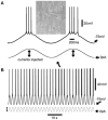
Figure 1
Figure 1
Sinusoidal current injection to induce cortical neuron up/down-states as slow-wave oscillations (SWOs). (A) Shows the two consecutive up/down-states with action potentials riding at the peaks with an inset of differential interference contrast (DIC) layer V cortical neuron image, expanded from two consecutive up/down-states in (B) (with an arrow indicating). (B) Shows the 60-s episode of SWOs (0.5 Hz) with injected sinusoidal current waveforms to induce neuronal up/down-states as SWOs. Scale bars are indicated as labeled. The vertical lines with dual arrows in (A,B) indicate the injected sinusoidal current amplitudes for up- (to induce action potentials) and down-states (0 pA injection, unless stated in Sections “Method/Result”). The sinusoidal waves are not in the same scale in (A,B).
Figure 2
Figure 2
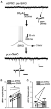
Artificial SWO induction potentiates spontaneous excitatory synaptic currents (sEPSCs) in layer V cortical neurons. (A) Shows one representative pre-SWO (top) and post-SWO sEPSC (below) in layer V cortical neurons and the middle panel shows one SWO. Individual sEPSC events (with arrow indicating) are expanded in a small temporal scale. Scale bars are indicated as labeled. (B) Shows the summary data of pre- (empty bar) and post-SWO (gray bar) sEPSC amplitudes from the same cortical neurons (dots connected with one line). (C) Shows the summary data of pre- (empty bar) and post-SWO (gray bar) sEPSC frequencies from the same cortical neurons (dots connected with one line). *Indicates a significant difference p < 0.05.
Figure 3
Figure 3
Artificial SWO induction potentiates spontaneous inhibitory synaptic currents (sIPSCs) in layer V cortical neurons. (A) One representative pre- (top) and post-SWO sIPSC (below) in layer V cortical neurons and the middle panel shows one SWO. Individual sIPSC events (with arrow indicating) are expanded in a small temporal scale. Scale bars are indicated as labeled. (B) Shows the summary data of pre- (empty bar) and post-SWO (gray bar) sIPSC amplitudes from the same cortical neurons (dots connected with one line). (C) Shows the summary data of pre- (empty bar) and post-SWO (gray bar) sIPSC frequencies from the same cortical neurons (dots connected with one line). *Indicates a significant difference p < 0.05.

References

    1. Abrahamsson T., Lalanne T., Watt A. J., Sjostrom J. P. (2016). Long-Term potentiation by theta-burst stimulation using extracellular field potential recordings in acute hippocampal slices. Cold Spring Harb. Protoc. 2016. 10.1101/pdb.prot091298 - DOI - PMC - PubMed
    1. Andrillon T., Nir Y., Staba R. J., Ferrarelli F., Cirelli C., Tononi G., et al. . (2011). Sleep spindles in humans: insights from intracranial EEG and unit recordings. J. Neurosci. 31, 17821–17834. 10.1523/JNEUROSCI.2604-11.2011 - DOI - PMC - PubMed
    1. Aoto J., Nam C. I., Poon M. M., Ting P., Chen L. (2008). Synaptic signaling by all-trans retinoic acid in homeostatic synaptic plasticity. Neuron 60, 308–320. 10.1016/j.neuron.2008.08.012 - DOI - PMC - PubMed
    1. Cantero J. L., Atienza M., Stickgold R., Kahana M. J., Madsen J. R., Kocsis B. (2003). Sleep-dependent theta oscillations in the human hippocampus and neocortex. J. Neurosci. 23, 10897–10903. 10.1523/JNEUROSCI.23-34-10897.2003 - DOI - PMC - PubMed
    1. Cataldi M., Panuccio G., Cavaccini A., D'Antuono M., Taglialatela M., Avoli M. (2011). Involvement of inward rectifier and M-type currents in carbachol-induced epileptiform synchronization. Neuropharmacology 60, 653–661. 10.1016/j.neuropharm.2010.11.023 - DOI - PMC - PubMed

LinkOut - more resources