Review
. 2022 Oct 14:10:1011631.
doi: 10.3389/fped.2022.1011631. eCollection 2022.

Comprehensive assessments of pulmonary circulation in children with pulmonary hypertension associated with congenital heart disease

Affiliations
Review

Comprehensive assessments of pulmonary circulation in children with pulmonary hypertension associated with congenital heart disease

Jun Muneuchi et al. Front Pediatr. .

Abstract

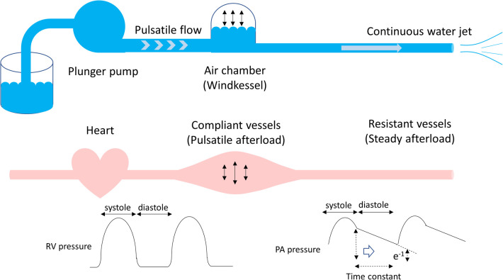
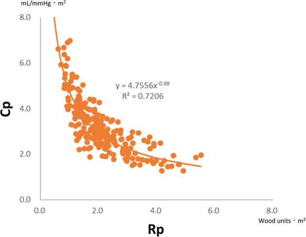
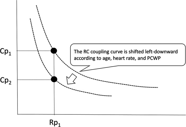
Pulmonary hypertension associated with congenital heart disease (CHD-PH) encompasses different conditions confounded by the left-to-right shunt, left heart obstruction, ventricular dysfunction, hypoxia due to airway obstruction, dysplasia/hypoplasia of the pulmonary vasculature, pulmonary vascular obstructive disease, and genetic variations of vasoactive mediators. Pulmonary input impedance consists of the pulmonary vascular resistance (Rp) and capacitance (Cp). Rp is calculated as the transpulmonary pressure divided by the pulmonary cardiac output, whereas Cp is calculated as the pulmonary stroke volume divided by the pulmonary arterial pulse pressure. The plots of Rp and Cp demonstrate a unique hyperbolic relationship, namely, the resistor-capacitor coupling curve, which represents the pulmonary vascular condition. The product of Rp and Cp is the exponential pressure decay, which refers to the time constant. Alterations in Cp are more considerable in CHD patients at an early stage of developing pulmonary hypertension or with excessive pulmonary blood flow due to a left-to-right shunt. The importance of Cp has gained attention because recent reports have shown that low Cp potentially reflects poor prognosis in patients with CHD-PH and idiopathic pulmonary hypertension. It is also known that Cp levels decrease in specific populations, such as preterm infants and trisomy 21. Therefore, both Rp and Cp should be individually evaluated in the management of children with CHD-PH who have different disease conditions.

Keywords: pulmonary arterial capacitance; pulmonary arterial compliance; pulmonary arterial hypertension; pulmonary vascular resistance; resistor–capacitor time; time constant (tau).


Conflict of interest statement

The authors declare that the research was conducted in the absence of any commercial or financial relationships that could be construed as a potential conflict of interest.

Figures

Figure 1
Figure 1
Windkessel model. The rhythmic water output of the plunger strokes is transformed into a continuous water jet downstream through an air tank. In terms of the pulmonary circulation, the plunger pump, an air tank, and a duct represent the heart, compliant vessels, and resistant vessels, respectively. The pulmonary arterial input impedance consists of compliant and resistant vessels, which determine the right ventricular afterload. The product of Rp and Cp is time-constant, which is an exponential pressure decay in diastole, referring time until pulmonary arterial pressure decreases to e−1 (37%) of pulmonary arterial end-systolic pressure. Rp, pulmonary vascular resistance; Cp, pulmonary vascular capacitance.
Figure 2
Figure 2
Cp determined by distention and recruitment of pulmonary vessels including capillaries. When cardiac output increases, the blood flow is accommodated by distention and recruitment, ensuring that the pulmonary circulation is highly compliant. Cp, pulmonary vascular capacitance.
Figure 3
Figure 3
Plots of Rp and Cp in 200 infants with ventricular septal defect and pulmonary hypertension (mean pulmonary arterial pressure >20 mmHg). There is a universal hyperbolic relationship between Rp and Cp. Rp, pulmonary vascular resistance; Cp, pulmonary vascular capacitance.
Figure 4
Figure 4
Unique relationship between Rp and Cp. A patient who is developing pulmonary hypertension is placed in the upper left region of the RC coupling curve (Patient 1, open circle) and moves from left to right along the curve. Then, a subtle increase in Rp is accompanied by a substantial decrease in Cp. Meanwhile, in a patent with more advanced stages of the disease (Patient 2, closed circle), Cp has already reached the minimum and any further increase in Rp accompanies no meaningful change in Cp. Rp, pulmonary vascular resistance; Cp, pulmonary vascular capacitance; RC, resistor–capacitor.
Figure 5
Figure 5
RC coupling among infants with ventricular septal defect and pulmonary hypertension; patients with excessively increased pulmonary blood flow are distributed in the upper left part of the RC coupling curve (open circle), whereas patients with modestly increased pulmonary blood flow are distributed in the lower right part of the curve (closed circle), which suggested that a subtle change in Rp gives a substantial change in Cp in patients with excessive pulmonary blood flow. Rp, pulmonary vascular resistance; Cp, pulmonary vascular capacitance; RC, resistor–capacitor.
Figure 6
Figure 6
RC coupling curve can be shifted left-downward because the RC time can be altered according to age, heart rate, and pulmonary capillary wedge pressure. When the RC coupling curve is shifted, one value of Rp gives different values of Cp. Rp, pulmonary vascular resistance; Cp, pulmonary vascular capacitance; RC, resistor–capacitor.

Similar articles

Cited by

References

    1. Astrand PO, Cuddy TE, Saltin B, Stenberg J. Cardiac output during submaximal and maximal work. J Appl Physiol. (1964) 19:268–74. 10.1152/jappl.1964.19.2.268 - DOI - PubMed
    1. Kovacs G, Berghold A, Scheidl S, Olschewski H. Pulmonary arterial pressure during rest and exercise in healthy subjects: a systematic review. Eur Respir J. (2009) 34:888–94. 10.1183/09031936.00145608 - DOI - PubMed
    1. Beshay S, Sahay S, Humbert M. Evaluation and management of pulmonary arterial hypertension. Respir Med. (2020) 171:106099. 10.1016/j.rmed.2020.106099 - DOI - PubMed
    1. Galiè N, Hoeper MM, Humbert M, Torbicki A, Vachiery JL, Barbera JA, et al. Guidelines for the diagnosis and treatment of pulmonary hypertension: the Task Force for the Diagnosis and Treatment of Pulmonary Hypertension of the European Society of Cardiology (ESC) and the European Respiratory Society (ERS), endorsed by the International Society of Heart and Lung Transplantation (ISHLT). Eur Heart J. (2009) 30:2493–537. 10.1093/eurheartj/ehp297 - DOI - PubMed
    1. Frank O. The basic shape of the arterial pulse. First treatise: mathematical analysis. 1899. J Mol Cell Cardiol. (1990) 22:255–77. 10.1016/0022-2828(90)91460-o - DOI - PubMed