Review
. 2023 Feb;115(2):e2200073.
doi: 10.1111/boc.202200073. Epub 2022 Nov 14.

A snapshot of protein trafficking in SARS-CoV-2 infection

Affiliations
Review

A snapshot of protein trafficking in SARS-CoV-2 infection

Vibhu Prasad et al. Biol Cell. 2023 Feb.

Abstract

SARS-CoV-2 is a human pathogenic virus responsible for the COVID-19 (coronavirus disease 2019) pandemic. The infection cycle of SARS-CoV-2 involves several related steps, including virus entry, gene expression, RNA replication, assembly of infectious virions and their egress. For all of these steps, the virus relies on and exploits host cell factors, cellular organelles, and processes such as endocytosis, nuclear transport, protein secretion, metabolite transport at membrane contact sites (MSC) and exocytotic pathways. To do this, SARS-CoV-2 has evolved multifunctional viral proteins that hijack cellular factors and modulate their function by unique strategies. In this Review, we highlight cellular trafficking factors, processes, and organelles of relevance to the SARS-CoV-2 infection cycle and how viral proteins make use of and perturb cellular transport during the viral infection cycle.


Conflict of interest statement

None declared.

Figures
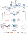
FIGURE 1
FIGURE 1
SARS‐CoV‐2 entry and endocytic trafficking. SARS‐CoV‐2 binds to cell surface receptor ACE2 and gets internalised in endosomes. Low pH endosomal environment and host proteases mediate viral fusion in the endocytic route (top left). Signals like endosomal pH, ions, and host cell factors (proteins and the lipid cholesterol) promote membrane fusion and uncoating of the SARS‐CoV‐2 genome. Non‐endocytic entry of SARS‐CoV‐2 (top right) takes place by the action of the TMPRSS2 protease, exposing membrane fusion peptide S2’ in S1/S2 cleaved spike. Binding of NRP1 also promotes the entry of SARS‐CoV‐2. Several pharmacological compounds inhibit individual steps in endocytic and non‐endocytic route of SARS‐CoV‐2 entry.
FIGURE 2
FIGURE 2
Inhibition of nucleo‐cytoplasmic trafficking of host mRNA in SARS‐CoV‐2 infection. (a) SARS‐CoV‐2 nsp1 binds to the nuclear export receptor heterodimer NXF1‐NXT1 preventing its association with nucleoporins and perturbing its docking to the nuclear pore complex. (b) ORF6 protein of SARS‐CoV‐2 interacts with nuclear pore complex components Rae1‐Nup98 causing their dislocation from the nuclear pore complex. ORF6 bound Rae1‐Nup98 was found predominantly in the cytoplasm leading to accumulation of cellular mRNAs in the nucleus. (c) SARS‐CoV‐2 nsp16 binds to the U1 and U2 snRNAs (small nuclear RNAs) preventing their association with the pre mRNA, there by inhibiting the maturation of cellular mRNA in the nucleus.
FIGURE 3
FIGURE 3
Secretion and exosomal trafficking in SARS‐CoV‐2 infection. (a) SARS‐CoV‐2 nsp8 and nsp9 block secretion by binding to the 7SL RNA of the SRP. Displacement of SRP by nsp8/9 leads to failure of nascent peptide‐to‐ER translocation, protein folding and secretion. Numbers indicate the progression of events from (1) identification of signal sequence (SS) on the mRNA by SRP complex; (2) assembly of the SRP‐ribosome complex on the translocon; (3) cleaving of the SS and co‐translational translocation; (4) protein folding in the ER lumen; and (5) vesicular transport. (b) Intracellular trafficking regulating ACE2 secretion pathways (left and right half of the figure, respectively) and ACE2 cell surface abundance (right panel). ACE2 is internalised after SARS‐CoV‐2 binding and is recycled to the cell surface via the endocytic route. Berbamine blocks TRPML‐Ca2+ channels at the lysosomal membrane, leading to inhibition of endolysosomal trafficking of ACE2 and resulting in lower ACE2 cell surface abundance and increased secretion via the exosomal pathway. Cell surface ACE2 can be taken up in the multi‐vesicular body and secreted as extra‐vesicular ACE2 (evACE2), which can potentially bind to and block the extracellular SARS‐CoV‐2 particles. Newly synthesised ACE2 in the ER traveling to the cell surface in secretory vesicles can be cleaved by ADAM17 and secreted from cells as soluble ACE2 (sACE2). (c) SARS‐CoV‐2 Orf3a inhibits the maturation of autophagosomes by blocking autophagosome‐lysosome fusion. Orf3a binds to the HOPS complex and inhibits HOPS‐SNAP29‐STX17 trimeric complex formation and autophagosome‐lysosome fusion. Numbers show the progression of events in virus infection cycle from (1) virus binding and internalisation; (2) RNA transcription and replication; (3) translation of sub‐genomic mRNAs to produce structural and accessory proteins; (4) inhibition of lysosome autophagosome fusion by Orf3a via interaction with HOPS; (5) inhibition of autophagosmal maturation and viral replication in the DMVs.
FIGURE 4
FIGURE 4
Membrane contact sites (MCS) at the PM ‐ ER interface in SARS‐CoV‐2 infection. ORAI1‐STIM1 regulated MCS at the PM ‐ ER interface regulate cytoplasmic Ca2+ dependent IFN‐I signalling response. (1) internalisation of Ca2+ ions from extracellular space to ER lumen via ORAI1‐STIM1 establishes MCSs between PM and the ER; (2) movement of Ca2+ ions from ER to cytoplasm via IP3R Ca2+ channels; (3) Ca2+‐dependent activation of NF‐AT and NF‐kB and transcriptional upregulation of interferon and inflammatory cytokine encoding genes; (4) expression and secretion of IFNs and inflammatory cytokines; (5) inhibition of SARS‐CoV‐2 infection by released cytokines.
FIGURE 5
FIGURE 5
Membrane contact sites (MCS) at Golgi ‐ endosome interface and ER‐derived replication organelle in SARS‐CoV‐2 infection. (a) Hybrid pre‐autophagosomal structure (HyPAS) formed by fusion of closely apposed FIP200‐positive cis‐Golgi‐derived vesicles and ATG16L1‐positive endosomes. HyPAS generated autophagosomes contribute to mito‐, lipo‐ and bulk‐autophagy. Formation of HyPAS depends on the SNARE STX17 and its interaction partners SIGMAR1, SERCA2, and E‐SYT2. HyPAS formation is affected by Ca2+ and SARS‐CoV‐2 nsp6 inhibits HyPAS by interacting with E‐SYT2, VAMP7, SIGMAR1 and SERCA2, hence disrupting Ca2+ regulation. (b) MCS between ER, SARS‐CoV‐2 induced DMVs, and lipid droplets generated by nsp6‐mediated ER membrane zippering. Zoomed in region shows selective occlusion of the ER lumen and membrane proteins as well as regulated lipid transfer via recruitment of the DFCP1‐Rab18 complex at the nsp6‐zippered ER membrane region.

References

    1. Abdelrahman, Z. , Li, M. & Wang, X. (2020) Comparative review of SARS‐CoV‐2, SARS‐CoV, MERS‐CoV, and influenza A respiratory viruses. Frontiers in Immunology, 11, 552909. - PMC - PubMed
    1. Addetia, A. , Lieberman, N.A.P. , Phung, Q. , Hsiang, T.Y. , Xie, H. , Roychoudhury, P. , Shrestha, L. , Loprieno, M.A. , Huang, M.‐L. , Gale, M. , Jerome, K.R. & Greninger, A.L. (2021) SARS‐CoV‐2 ORF6 disrupts bidirectional nucleocytoplasmic transport through interactions with Rae1 and Nup98. mBio, 12; e0065‐21. - PMC - PubMed
    1. Akopian, D. , Shen, K. , Zhang, X. & Shan, S.‐O. (2013) Signal recognition particle: an essential protein‐targeting machine. Annual Review of Biochemistry, 82, 693–721. - PMC - PubMed
    1. Badawi, S. & Ali, BR. (2021) ACE2 Nascence, trafficking, and SARS‐CoV‐2 pathogenesis: the saga continues. Human Genomics, 15, 8. - PMC - PubMed
    1. Banerjee, A.K. , Blanco, M.R. , Bruce, E.A. , Honson, D.D. , Chen, L.M. , Chow, A. , Bhat, P. , Ollikainen, N. , Quinodoz, S.A. , Loney, C. , Thai, J. , Miller, Z.D. , Lin, A.E. , Schmidt, M.M. , Stewart, D.G. , Goldfarb, D. , De Lorenzo, G. , Rihn, S.J. , Voorhees, R.M. , Botten, J.W. , Majumdar, D. & Guttman, M. (2020) SARS‐CoV‐2 disrupts splicing, translation, and protein trafficking to suppress host defenses. Cell, 183, 1325‐1339.e21 e1321. - PMC - PubMed