. 2022 Dec 20;13(6):e0221822.
doi: 10.1128/mbio.02218-22. Epub 2022 Oct 31.

The Paxillin MoPax1 Activates Mitogen-Activated Protein (MAP) Kinase Signaling Pathways and Autophagy through MAP Kinase Activator MoMka1 during Appressorium-Mediated Plant Infection by the Rice Blast Fungus Magnaporthe oryzae

Affiliations

The Paxillin MoPax1 Activates Mitogen-Activated Protein (MAP) Kinase Signaling Pathways and Autophagy through MAP Kinase Activator MoMka1 during Appressorium-Mediated Plant Infection by the Rice Blast Fungus Magnaporthe oryzae

Wuyun Lv et al. mBio. .

Abstract

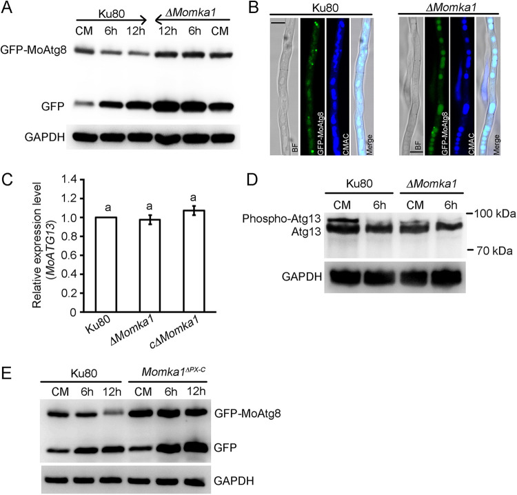
Paxillin is a focal adhesion-associated protein that functions as an adaptor to recruit diverse cytoskeleton and signaling molecules into a complex and plays a crucial role in several signaling pathways in mammal cells. However, paxillin-mediated signal pathways are largely unknown in phytopathogenic fungi. Previously, Pax1 of Magnaporthe oryzae (MoPax1), a paxillin-like protein, has been identified as a crucial pathogenicity determinant. Here, we report the identification of a mitogen-activated protein (MAP) kinase (MAPK) activator, Mka1 of M. oryzae (MoMka1), that physically interacts with MoPax1. Targeted gene deletion of MoMKA1 resulted in pleiotropic defects in aerial hyphal growth, conidiation, appressorium formation, and pathogenicity in M. oryzae. MoMka1 interacts with Mst50, an adaptor protein of the Mst11-Mst7-Pmk1 and Mck1-Mkk2-Mps1 cascades. Moreover, the phosphorylation levels of both Pmk1 and Mps1 in aerial hyphae of the ΔMomka1 mutant were significantly reduced, indicating that MoMka1 acts upstream from the MAPK pathways. Interestingly, we found that MoMka1 interacts with MoAtg6 and MoAtg13. Deletion of MoMKA1 led to impaired MoAtg13 phosphorylation and enhanced autophagic flux under nutrient-rich conditions, indicating that MoMka1 is required for regulation of autophagy in M. oryzae. Taken together, the paxillin MoPax1 may activate MAP kinase signaling pathways and autophagy through MAP kinase activator MoMka1 and play important roles during appressorium-mediated plant infection by the rice blast fungus. IMPORTANCE Paxillin, as an adaptor recruiting diverse cytoskeleton and signaling molecules into a complex, plays a crucial role in several signaling pathways in mammal cells. However, paxillin-mediated signal pathways are largely unknown in phytopathogenic fungi. Here, we identified that MoMka1 physically interacts with MoPax1. Furthermore, MoMka1 acts upstream from the MAPK pathways through interacting with Mst50, a key protein of the Mst11-Mst7-Pmk1 and Mck1-Mkk2-Mps1 cascades. Meanwhile, MoMka1 interacts with both MoAtg6 and MoAtg13 and controls autophagy initiation by influencing the phosphorylation level of MoAtg13. In summary, we describe a model in which MoPax1 activates MAP kinase signaling pathways and autophagy through MoMka1 during appressorium-mediated plant infection by M. oryzae.

Keywords: Magnaporthe oryzae; MoMka1; MoPax1; appressoria; autophagy; mitogen-activated protein kinases.


Conflict of interest statement

The authors declare no conflict of interest.

Figures

FIG 1
FIG 1
MoMka1 interacts with MoPax1 via the second SH3 domain in M. oryzae. (A) Yeast two-hybrid (Y2H) analysis of the interaction between MoMka1 and MoPax1. The fusion constructs MoMKA1-pGADT7 and MoPAX1-pGBKT7 were cointroduced into the yeast Y2H Gold strain, and transformants were cultured on synthetic defined medium (SD)-Leu-Trp plates as a control and on SD-Ade-Leu-Trp-His for 3 days. The pair of plasmids pGADT7-T and pGBKT7-53 was used as the positive control, and the pair of plasmids pGADT7-T and pGBKT7-Lam was used as the negative control. AD, the pGADT7 plasmid; BD, the pGBKT7 plasmid. (B) Coimmunoprecipitation (co-IP) assays for the interaction between MoMka1 and MoPax1. The MoMka1-GFP and MoPax1-3×FLAG fusion constructs were coexpressed in the Ku80 strain. The isolated proteins were analyzed by Western blotting using anti-GFP and anti-Flag antibodies. (C) Domain map of MoMka1. The domain prediction of MoMka1 was performed with the SMART analysis service. (D) Y2H assays between four MoMka1 variants and MoPax1. I, only containing the first SH3 domain; II, carrying two SH3 domains; III, lacking the first SH3 domain; IV, lacking the PB1 domain; Ⅴ, only containing the second SH3 domain; Ⅵ, lacking the second SH3 domain. Pairs of different combinations of the truncated constructs of MoMka1 and MoPax1 were cotransformed into Y2H Gold.
FIG 2
FIG 2
The phenotype analysis of the ΔMomka1 mutant. (A) Colonies of the Ku80 strain, the ΔMomka1 mutant, and the cΔMomka1 complemented strain cultured on CM plates at 25°C for 10 days. (B) Radial growth of each strain on CM plates. Error bars represent standard deviations. The same small letter indicates no significant difference (P < 0.05). (C) Bar chart showing statistical analysis of conidiation. Each strain was cultured on CM plates at 25°C for 12 days. Error bars represent standard deviations. **, P < 0.01. (D) Microscopic observation of conidial development. Conidial formation of each strain was observed under a light microscope after incubation on coverslips at 28°C for 24 h. Bar = 100 μm. (E) Microscopic observation of conidial morphology. Conidia of each strain were collected from the CM plates after growth at 25°C for 12 days. Bar = 10 μm.
FIG 3
FIG 3
MoMKA1 is involved in appressorium formation in M. oryzae. (A) Microscopic observation of appressorium development of the Ku80 strain, the ΔMomka1 mutant, and the cΔMomka1 complemented strain. Conidia of each strain were harvested from the CM plates and allowed to form appressoria on hydrophobic surfaces (24 h). Bar = 10 μm. (B) Statistical analysis of appressorium formation rates. Error bars represent standard deviations. **, P < 0.01. (C) Quantification of collapsed appressoria of each strain. For each glycerol concentration, at least 100 appressoria were observed. Error bars represent standard deviations. *, P < 0.05. (D) The pattern of expression of MoMKA1 during different stages of development by qRT-PCR analysis. MY, mycelium. *, P < 0.05; **, P < 0.01.
FIG 4
FIG 4
MoMKA1 is essential for pathogenicity in M. oryzae. (A) Barley cut-leaf assays. With 20 μL per drop, conidial suspensions (5 × 104 conidia/mL) of each strain were inoculated onto detached barley leaves. Photographs were taken at 5 dpi. (B) Spray inoculation assays. Rice seedlings were spray inoculated with 10 mL conidial suspension (2 × 104 conidia/mL) of each strain. Photographs were taken at 5 dpi. (C) Penetration assays in the epidermis of barley leaves. Conidial suspensions (5 × 104 conidia/mL) of each strain were drop inoculated onto the underside of detached barley leaves. After 24 h at 28°C in dark, the epidermis of barley leaves was torn and observed under a light microscope. AP, appressorium; IH, invasion hyphae. Bar = 20 μm. (D) Statistical analysis of penetration and invasion growth on onion epidermis cells. Error bars represent standard deviations. Type 1, invasion hyphae with branches; type 2, invasion hyphae with a single branch; type 3, no penetration; type 4, no germination. AP, appressorium; IH, invasion hyphae; CO, conidia. Bar = 20 μm.
FIG 5
FIG 5
Subcellular localization of MoMka1 in hyphae (A), conidia (B), and mature appressoria (C) of the cΔMomka1 complemented strain. GFP fluorescence was examined under fluorescence confocal microscopy. Arrowheads point to punctate green fluorescence. BF, bright-field microscopy. Bar = 5 μm.
FIG 6
FIG 6
MoMka1 interacts with Mst50 and is involved in MAPK signaling pathways in M. oryzae. (A) Y2H analysis of the interaction between MoMka1 and Mst50. The fusion constructs MoMKA1-pGADT7 and MST50-pGBKT7 were cointroduced into the yeast Y2H Gold strain, and transformants were cultured on SD-Leu-Trp plates as a control and on SD-Ade-Leu-Trp-His for 3 days. The pair of plasmids pGADT7-T and pGBKT7-53 was used as the positive control, and the pair of plasmids pGADT7-T and pGBKT7-Lam was used as the negative control. (B) Co-IP assays for the interaction between MoMka1 and Mst50. Total proteins (input) extracted from the strains containing the Mst50-3×FLAG and MoMka1-GFP constructs or a single construct (Mst50-3×FLAG or MoMka1-GFP) were subjected to SDS-PAGE, and immunoblots were incubated with anti-FLAG and anti-GFP antibodies. In addition, each protein sample was pulled down using anti-FLAG antibody and further detected with anti-GFP antibody (IP). The protein samples were also detected with anti-GAPDH antibody as a reference. (C) TpEY phosphorylation assays. Total proteins were isolated from mycelia of each strain. The anti-Pmk1 antiserum detected a 42-kDa Pmk1 band in all the samples. An anti-TpEY antibody detected the phosphorylation levels of Pmk1 and Mps1 (46 kDa). (D) Conidial germination assays. Conidial suspensions of each strain were inoculated onto hydrophobic surfaces with or without 50 μg/mL calcofluor white (CFW). Mean values and standard deviations of the germination rates assayed at 6 h were calculated from three replicates. Error bars represent standard deviations. **, P < 0.01. (E) The relative expression levels of MoMKA1 in CM liquid medium with or without CFW by qRT-PCR analysis. Error bars represent standard deviations. **, P < 0.01.
FIG 7
FIG 7
MoMka1 interacts with MoAtg13 and MoAtg6. (A) The Y2H assay of interaction between MoMka1 and MoAtg13. Serial concentrations of yeast cells transformed with bait and prey constructs indicated in the figure were assayed for growth on SD-Ade-Leu-Trp-His for 3 days. (B) Y2H analysis of the interaction between MoMka1 and MoAtg6. (C) Co-IP assays for the interaction between MoMka1 and MoAtg13. Total proteins (input) extracted from the strains containing the MoMka1-GFP and MoAtg13-3×FLAG constructs or a single construct (MoMka1-GFP or MoAtg13-3×FLAG) were probed with anti-GFP and anti-FLAG antibodies. In addition, each protein sample was pulled down using anti-FLAG antibody and further detected with anti-GFP antibody (IP). The protein samples were also detected with anti-GAPDH antibody as a reference. (D) Co-IP assays for the interaction between MoMka1 and MoAtg6.
FIG 8
FIG 8
MoMKA1 regulates autophagy through affecting the phosphorylation level of MoAtg13 in M. oryzae. (A) GFP-MoAtg8 proteolysis assays of the Ku80 strain and the ΔMomka1 mutant expressing the GFP-MoAtg8 fusion construct. Total proteins were extracted for analysis by Western blotting with anti-GFP antibody. Anti-GAPDH antibody was used as a control. (B) GFP-MoAtg8 localization in the hyphal cells of Ku80 and the ΔMomka1 mutant under nutrient-rich conditions (CM medium). The vacuoles were stained with CMAC (7-amino-4-chloromethycoumarin) and examined by fluorescence microscopy. Bar = 5 μm. (C) The expression level of MoATG13 in each strain cultured in CM medium as determined by qRT-PCR analysis. Error bars represent standard deviations. The same small letter indicates no significant difference (P < 0.05). (D) Detection of the MoAtg13 phosphorylation level in Ku80 and the ΔMomka1 mutant. Lysates derived from Ku80 and the ΔMomka1 mutant cultured in CM liquid medium and MM-N medium for 6 h were subjected to SDS-PAGE and immunoblotting with anti-Atg13 antibody. Anti-GAPDH antibody was also used as a control. (E) GFP-MoAtg8 proteolysis assays of Ku80 and the Momka1ΔPX-C mutant with the PX domain deleted. Total proteins were extracted for analysis by Western blotting with anti-GFP antibody. Anti-GAPDH antibody was used as a control.
FIG 9
FIG 9
A model in which MoPax1 activates MAP kinase signaling pathways and autophagy through MoMka1 during appressorium-mediated plant infection by M. oryzae.

Similar articles

Cited by

References

    1. Wilson RA, Talbot NJ. 2009. Under pressure: investigating the biology of plant infection by Magnaporthe oryzae. Nat Rev Microbiol 7:185–195. doi:10.1038/nrmicro2032. - DOI - PubMed
    1. Callaway E. 2016. Devastating wheat fungus appears in Asia for first time. Nature 532:421–422. doi:10.1038/532421a. - DOI - PubMed
    1. Tucker SL, Talbot NJ. 2001. Surface attachment and pre-penetration stage development by plant pathogenic fungi. Annu Rev Phytopathol 39:385–417. doi:10.1146/annurev.phyto.39.1.385. - DOI - PubMed
    1. De Jong JC, McCormack BJ, Smirnoff N, Talbot NJ. 1997. Glycerol generates turgor in rice blast. Nature 389:244–244. doi:10.1038/38418. - DOI
    1. Talbot NJ. 1995. Having a blast: exploring the pathogenicity of Magnaporthe grisea. Trends Microbiol 3:9–16. doi:10.1016/S0966-842X(00)88862-9. - DOI - PubMed

Publication types

Supplementary concepts