. 2022 Nov 23;96(22):e0099722.
doi: 10.1128/jvi.00997-22. Epub 2022 Oct 31.

WTAP Targets the METTL3 m6A-Methyltransferase Complex to Cytoplasmic Hepatitis C Virus RNA to Regulate Infection

Affiliations

WTAP Targets the METTL3 m6A-Methyltransferase Complex to Cytoplasmic Hepatitis C Virus RNA to Regulate Infection

Matthew T Sacco et al. J Virol. .

Abstract

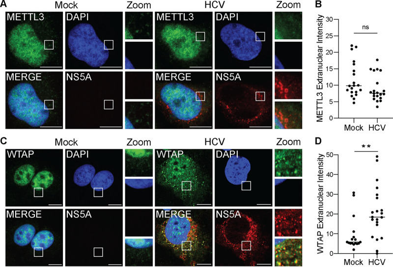
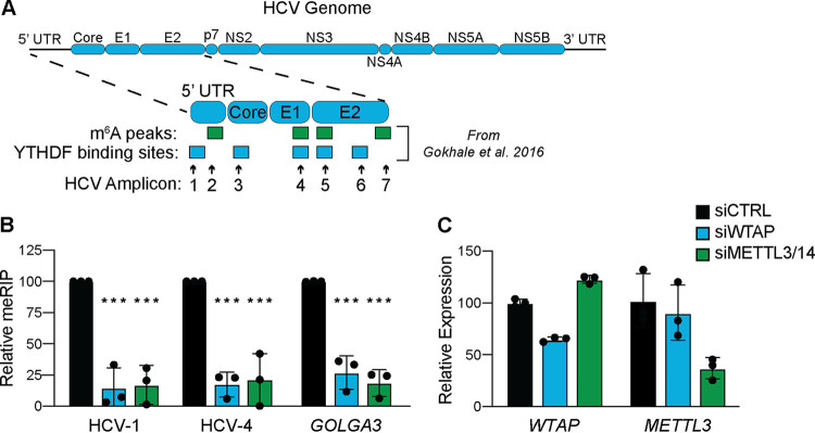
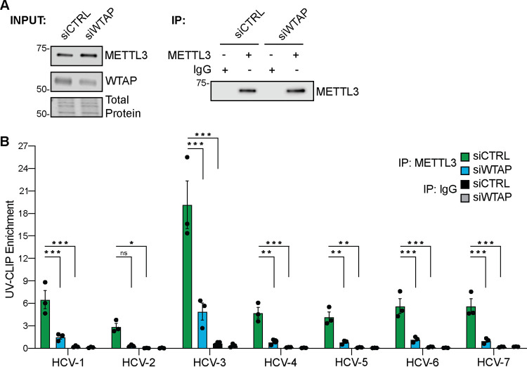
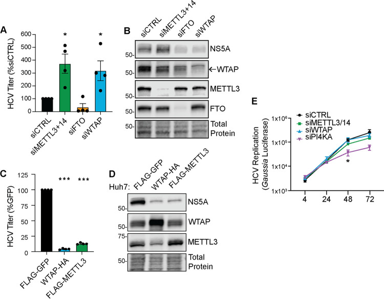
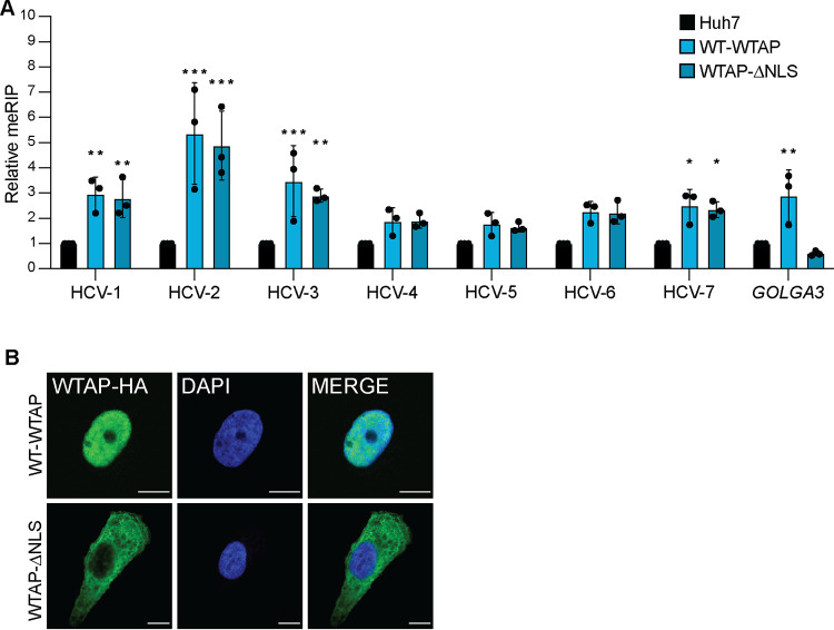
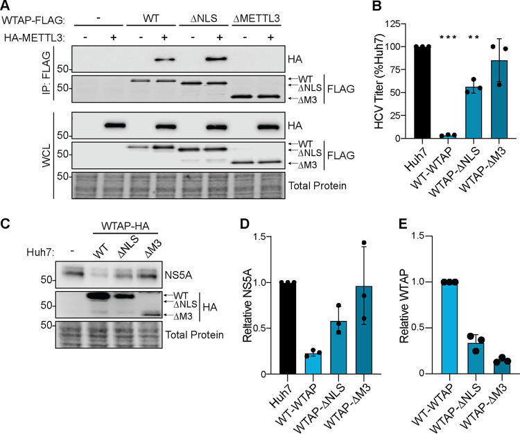
Modification of the hepatitis C virus (HCV) positive-strand RNA genome by N6-methyladenosine (m6A) regulates the viral life cycle. This life cycle takes place solely in the cytoplasm, while m6A addition on cellular mRNA takes place in the nucleus. Thus, the mechanisms by which m6A is deposited on the viral RNA have been unclear. In this work, we find that m6A modification of HCV RNA by the m6A-methyltransferase proteins methyltransferase-like 3 and 14 (METTL3 and METTL14) is regulated by Wilms' tumor 1-associating protein (WTAP). WTAP, a predominantly nuclear protein, is an essential member of the cellular mRNA m6A-methyltransferase complex and known to target METTL3 to mRNA. We found that HCV infection induces localization of WTAP to the cytoplasm. Importantly, we found that WTAP is required for both METTL3 interaction with HCV RNA and m6A modification across the viral RNA genome. Further, we found that WTAP, like METTL3 and METTL14, negatively regulates the production of infectious HCV virions, a process that we have previously shown is regulated by m6A. Excitingly, WTAP regulation of both HCV RNA m6A modification and virion production was independent of its ability to localize to the nucleus. Together, these results reveal that WTAP is critical for HCV RNA m6A modification by METTL3 and METTL14 in the cytoplasm. IMPORTANCE Positive-strand RNA viruses such as HCV represent a significant global health burden. Previous work has described that HCV RNA contains the RNA modification m6A and how this modification regulates viral infection. Yet, how this modification is targeted to HCV RNA has remained unclear due to the incompatibility of the nuclear cellular processes that drive m6A modification with the cytoplasmic HCV life cycle. In this study, we present evidence for how m6A modification is targeted to HCV RNA in the cytoplasm by a mechanism in which WTAP recruits the m6A-methyltransferase METTL3 to HCV RNA. This targeting strategy for m6A modification of cytoplasmic RNA viruses is likely relevant for other m6A-modified positive-strand RNA viruses with cytoplasmic life cycles such as enterovirus 71 and SARS-CoV-2 and provides an exciting new target for potential antiviral therapies.

Keywords: HCV; METTL14; METTL3; N6-methyladenosine; RNA modification; WTAP.


Conflict of interest statement

The authors declare no conflict of interest.

Figures

FIG 1
FIG 1
HCV infection alters the subcellular localization of the m6A accessory protein WTAP. (A and C) Confocal micrographs of mock- or HCV (48 h, MOI of 0.3)-infected Huh7 cells stained with DAPI and antibodies against HCV NS5A and either METTL3 (A) or WTAP (C). Zoom is taken from area in the white box. (B and D) Quantification of the fluorescent signal intensity in the extranuclear region of Huh7 cells for METTL3 (B) or WTAP (D), as described in Materials and Methods for fields of mock (NS5A-negative)- or HCV-infected (NS5A-positive) cells. Scale bars = 10 μM. Graph shows mean ± SD; n = 21 fields. Data were analyzed by Welch’s unequal variances t test (**, P < 0.01; ns, not significant).
FIG 2
FIG 2
WTAP and METTL3 + 14 are essential for m6A modification of HCV RNA. (A) Illustration of the HCV RNA genome with amplicons measured in this study and m6A peaks or YTHDF protein binding sites identified in reference . UTR, untranslated region. (B and C) Relative meRIP enrichment of the indicated viral or cellular amplicons from Huh7 cells treated with the indicated siRNAs and infected with HCV (48 h, MOI of 0. 3) (B), as determined as the percentage of input under each condition normalized to an m6A RNA spike-in with siCTRL samples normalized to 100%, and RT-qPCR analysis of indicated genes (C), relative to 18S rRNA. For panel B, graph shows mean ± SD (n = 3 biological replicates), while panel C is representative. Data were analyzed by two-way ANOVA with Šidák’s multiple-comparison test (***, P < 0.001).
FIG 3
FIG 3
METTL3 directly binds HCV RNA in a WTAP-dependent manner. Huh7 cells were treated with the indicated siRNA and infected with HCV (72 h, MOI 1), followed by UV-CLIP with anti-METTL3 or nonspecific IgG. (A) Immunoblot analysis of input and immunoprecipitated UV-CLIP lysates. (B) Enrichment of indicated amplicons by METTL3 or IgG immunoprecipitation, relative to input, as measured by RT-qPCR. Graph shows mean ± SD; n = 3 biological replicates. Blot is representative of 3 independent experiments. Data were analyzed by two-way ANOVA with Šidák’s multiple-comparison test (*, P < 0.05; **, P < 0.01; ***, P < 0.001; ns, not significant).
FIG 4
FIG 4
WTAP negatively regulates HCV virion production. (A and B) Focus-forming assay of supernatant harvested from HCV-infected (48 h, MOI of 0.3) Huh7 cells depleted of indicated proteins (A), as well as immunoblot analysis of these lysates (B). (C and D) Focus-forming assay of supernatant harvested from HCV-infected (48 h, MOI of 0.3) Huh7 cells overexpressing the indicated proteins (C), as well as immunoblot analysis of these lysates (D). For WTAP and METTL3, protein-specific antibodies detect both endogenous and overexpressed proteins. (E) Gaussia luciferase values from the supernatant of Huh7.5-CD81 KO cells treated with the indicated siRNA and transfected with a full-length HCV RNA containing a Gaussia luciferase reporter cassette, measured at indicated hour posttransfection. Graphs show mean ± SD; n = 3 (C and E) or 4 (A) biological replicates. Blots are representative of 3 independent experiments. Data were analyzed by one-way ANOVA with Šidák’s multiple-comparison test (*, P < 0.05; ***, P < 0.001).
FIG 5
FIG 5
WTAP regulation of HCV RNA m6A modification is independent of its nuclear localization. (A) Relative meRIP enrichment of indicated amplicons in HCV-infected (72 h, MOI of 1) parental Huh7-, wild-type (WT)-WTAP-HA-, or WTAPΔNLS-HA-overexpressing cells. (B) Confocal micrographs of Huh7 cells overexpressing WT-WTAP-HA or WTAPΔNLS-HA stained as indicated. Graph show mean ± SD; n = 3 biological replicates for panel A with micrographs of localization shown in panel B. Scale bars = 10 μM. Data were analyzed by two-way ANOVA with Šidák’s multiple-comparison test (*, P < 0.05; **, P < 0.01; ***, P < 0.001).
FIG 6
FIG 6
WTAP regulation of HCV virion production requires METTL3 interaction but not nuclear localization. (A) Immunoblot analysis of anti-FLAG-immunoprecipitated lysates from Huh7 cells cotransfected with HA-METTL3 and indicated WTAP-FLAG constructs, either wild-type, ΔNLS, or ΔMETTL3 interaction. (B to E) Focus-forming assay of supernatant harvested from HCV-infected (48 h, MOI of 0.3) Huh7 cells overexpressing the indicated protein (B), as well as immunoblot analysis of lysates (C) and quantification of NS5A and WTAP levels and relative total protein (D and E). Graphs show mean ± SD; n = 3 biological replicates. Blots are representative of 3 independent experiments. Data were analyzed by one-way ANOVA with Šidák’s multiple-comparison test (**, P < 0.01; ***, P < 0.001).

Update of

References

    1. Manns MP, Buti M, Gane E, Pawlotsky JM, Razavi H, Terrault N, Younossi Z. 2017. Hepatitis C virus infection. Nat Rev Dis Primers 3:17006. 10.1038/nrdp.2017.6. - DOI - PubMed
    1. Moradpour D, Penin F, Rice CM. 2007. Replication of hepatitis C virus. Nat Rev Microbiol 5:453–463. 10.1038/nrmicro1645. - DOI - PubMed
    1. Shulla A, Randall G. 2015. Spatiotemporal analysis of hepatitis C virus infection. PLoS Pathog 11:e1004758. 10.1371/journal.ppat.1004758. - DOI - PMC - PubMed
    1. Niepmann M, Shalamova LA, Gerresheim GK, Rossbach O. 2018. Signals involved in regulation of hepatitis C virus RNA genome translation and replication. Front Microbiol 9:395. 10.3389/fmicb.2018.00395. - DOI - PMC - PubMed
    1. Gokhale NS, McIntyre ABR, McFadden MJ, Roder AE, Kennedy EM, Gandara JA, Hopcraft SE, Quicke KM, Vazquez C, Willer J, Ilkayeva OR, Law BA, Holley CL, Garcia-Blanco MA, Evans MJ, Suthar MS, Bradrick SS, Mason CE, Horner SM. 2016. N6-methyladenosine in flaviviridae viral RNA genomes regulates infection. Cell Host Microbe 20:654–665. 10.1016/j.chom.2016.09.015. - DOI - PMC - PubMed

Publication types