. 2022 Nov 23;96(22):e0096322.
doi: 10.1128/jvi.00963-22. Epub 2022 Oct 31.

pUL36 Deubiquitinase Activity Augments Both the Initiation and the Progression of Lytic Herpes Simplex Virus Infection in IFN-Primed Cells

Affiliations

pUL36 Deubiquitinase Activity Augments Both the Initiation and the Progression of Lytic Herpes Simplex Virus Infection in IFN-Primed Cells

Jonas Mohnke et al. J Virol. .

Abstract

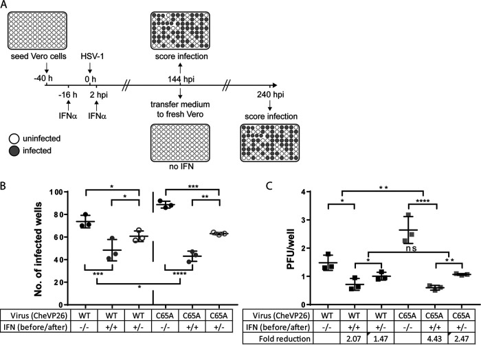
The evolutionarily conserved, structural HSV-1 tegument protein pUL36 is essential for both virus entry and assembly. While its N-terminal deubiquitinase (DUB) activity is dispensable for infection in cell culture, it is required for efficient virus spread in vivo, as it acts as a potent viral immune evasin. Interferon (IFN) induces the expression of hundreds of antiviral factors, including many ubiquitin modulators, which HSV-1 needs to neutralize to efficiently initiate a productive infection. Herein, we discover two functions of the conserved pUL36 DUB during lytic replication in cell culture in an understudied but equally important scenario of HSV-1 infection in IFN-treated cells. Our data indicate that the pUL36 DUB contributes to overcoming the IFN-mediated suppression of productive infection in both the early and late phases of HSV-1 infection. We show that incoming tegument-derived pUL36 DUB activity contributes to the IFN resistance of HSV-1 in IFN-primed cells to efficiently initiate lytic virus replication. Subsequently, the de novo expressed DUB augmented the efficiency of virus replication and increased the output of infectious virus. Notably, the DUB defect was only apparent when IFN was applied prior to infection. Our data indicate that IFN-induced defense mechanisms exist and that they work to both neutralize infectivity early on and slow the progression of HSV-1 replication in the late stages of infection. Also, our data indicate that pUL36 DUB activity contributes to the disarming of these host responses. IMPORTANCE HSV-1 is a ubiquitous human pathogen that is responsible for common cold sores and may also cause life-threatening disease. pUL36 is an essential, conserved herpesvirus protein with N-terminal deubiquitinating (DUB) activity. The DUB is dispensable for HSV-1 replication in cell culture but represents an important viral immune evasin in vivo. IFN plays a pivotal role in HSV-1 infection and suppresses viral replication both in vitro and in vivo. Here, we show that DUB activity contributes to overcoming IFN-induced cellular resistance in order to more efficiently initiate lytic replication and produce infectious virions. As such, DUB activity in the incoming virions increases their infectivity, while the de novo synthesized DUB augments productive infection. Thus, the HSV-1 DUB antagonizes the activity of IFN-inducible effector proteins to facilitate productive infection at multiple levels. Our findings underscore the importance of using more challenging cell culture systems to fully understand virus protein functions.

Keywords: DUB; HSV-1; UL36; USP; herpes simplex virus 1; innate immunity; interferon; interferon antagonism; ubiquitin.


Conflict of interest statement

The authors declare no conflict of interest.

Figures

FIG 1
FIG 1
Growth kinetics of parental HSV-1 and its pUL36 DUB mutants. (A) Multistep growth curves of the CheVP26 (black lines) and CheVP26-C65A (gray lines) viruses in untreated (no symbols), 500 IU/mL IFN-α pretreated (filled squares), or 500 IU/mL IFN-α and 100 IU/mL IFN-γ pretreated Vero cells (open triangles) (n = 3). IFN was added 20 h prior to inoculation and was replenished after inoculation. Cells were infected with a MOI of 0.001 (untreated and IFN-α) or 0.1 (IFN-α/γ) of either the parental CheVP26 or the CheVP26-C65A virus. The total virus yield was determined in 24 h intervals up to 120 hpi via plaque assay on IFN-naive Vero cells. (B) Plaque size measurements from KOS or KOS-C65A infected Vero cells. The results from a representative experiment are shown. Untreated or IFN pretreated (as in panel A) Vero cells were infected with ~100 PFU/well (100-fold more for IFN-α/γ) and fixed at 2 dpi for untreated cells and at 4 dpi for IFN-treated cells. Plaque sizes were measured using Fiji software. (A and B) Statistical analysis was performed using the Two-Way ANOVA analysis plug-in of GraphPad Prism. (C) Untreated or IFN pretreated Vero cells (500 IU/mL IFN-α alone or in combination with 100 IU/mL IFN-γ) were infected at a high MOI (untreated, MOI of 10; IFN-α, MOI of 20; IFN-α/γ, MOI of 100). The total virus (cells and medium) was harvested at 24 hpi, and the yield was determined via plaque assay on untreated Vero cells. The mean yield from two independent experiments is shown. (D) Untreated or IFN pretreated Vero cells (500 IU/mL IFN-α and 100 IU/mL IFN-γ) were infected for 1 h, and replicated viral DNA was measured at 6 hpi. The mean fold reduction of replication was calculated via the ΔΔCq method, using β-actin as a cellular reference. **, P < 0.01; ****, P < 0.0001.
FIG 2
FIG 2
Effect of the pUL36 DUB on plaque initiation. (A–D) Vero cells were either left untreated or pretreated with different regimens of IFN-α either alone (A–C) or in combination with IFN-γ (D). Plaque assays were performed 20 h after the treatment of the cells with IFN using ~100 PFU/well (A–D) or 10,000 PFU/well (panel D, only for the IFN-α/γ treated wells) of either parental or isogenic C65A mutants (strains indicated). The ratio of plaques that formed in the presence and absence of IFN (panels A–C, IFN-α/control) or the fold inhibition of plaque formation (D, control/IFN-α/γ) is shown. (B) Using the experimental setup employed in panel A with minor modifications (see Materials and Methods for the IFN-α concentrations used), the plaque formation was compared on untreated and IFN-α pretreated Vero and human SK-N-SH, HaCaT, and RPE-1 cells. (A–D) Statistical analysis was performed using the One-Way ANOVA analysis plug-in of GraphPad Prism. (E) Mass spectrometric analysis of three independently generated gradient purified virion preparations of the CheVP26 and CheVP26-C65A viruses (parental and mutant virus prepared as pairs on the same day). Differences in the identified virion components were compared between the different biological replicates (i.e., virion preparations) of the same (turquoise) and different (red) viruses. All abundances were normalized to VP5 content, and only viral proteins are shown. *, P < 0.05; **, P < 0.01; ns, not significant.
FIG 3
FIG 3
Effect of the DUB mutation on infectivity at high MOI. Untreated (no IFN) or IFN-α/γ pretreated Vero cells were infected with serially diluted inocula of either the CheVP26 or the CheVP26-C65A virus. MOIs of 10 and 1 are shown. The cells were fixed at 24 hpi and stained with anti-ICP4 antibody in order to detect infected cells. The cellular nuclei were stained with DAPI. Grayscale images with enhanced brightness and contrast are shown. Scale bars are indicated on the bottom right of each image. For clarification, a scale bar of 250 μm (identical for each image) was added underneath the figure.
FIG 4
FIG 4
The UL36 DUB augments plaque formation in IFN pretreated cells. (A) Vero cells, which were left untreated, pretreated with 100 or 500 IU/mL IFN-α for 20 h prior to inoculation only (“pre”), or treated with 100 or 500 IU/mL IFN-α prior to and after inoculation (“pre + post”), were infected with ~100 PFU/well of the KOS or KOS-C65A viruses. (B) Similar to panel A, except that 500 IU/mL IFN-α treatment was applied throughout the whole assay (“pre + post”) and was compared to treatment with 500 IU/mL IFN-α applied only after inoculation (“post”). (A and B) Plaque ratios of IFN-treated to IFN-untreated Vero cells were calculated and plotted. Statistical analysis was performed using the Two-Way ANOVA analysis plug-in of GraphPad Prism. *, P < 0.05; **, P < 0.01.
FIG 5
FIG 5
Tegument UL36 DUB activity augments plaque initiation in IFN pretreated cells. (A) Schematic representation of non-complemented DUB mutant virions (left panel) and complemented DUB mutant virions (right panel). When the C65A virus is grown on normal producer cells (those not expressing pUL36, e.g., RS cells) the virions, which are produced, only contain the mutant, virus-derived pUL36 protein (red). When the C65A virus is grown on UL36 complementing cells (e.g., RSC-HA-UL36), the resulting virions contain a mixture of mutant, virus-derived pUL36 and wild-type, cell-derived pUL36 (blue, right panel). Parental virus grown on the respective cells served as controls. After the initial infection of noncomplementing cells (e.g., Vero or SK-N-SH cells) with complemented DUB mutant, the plaques progressed as “mutant”, since the virions produced in these cells only contain de novo synthesized pUL36-C65A. (B and C) Vero or SK-N-SH cells were treated prior to and after inoculation with (B) 500 IU/mL IFN-α or (C) 25 IU/mL IFN-α and were infected at ~100 PFU/well with the indicated viruses. Complementation status is indicated as “C” (complemented) or “NC” (non-complemented). Plaque ratios were calculated and plotted as before. (D) Plaque sizes on untreated and IFN-α/γ pretreated Vero cells were analyzed for the complemented DUB mutant virus and compared to that of the non-complemented virus. Each data point represents one plaque which was normalized against the mean plaque size of the parental virus assayed simultaneously. (B–D) Statistical analysis was performed using the (C and D) One-Way ANOVA or (B) Two-Way ANOVA analysis plug-in of GraphPad Prism. *, P < 0.05; ***, P < 0.001; ****, P > 0.0001; ns, not significant.
FIG 6
FIG 6
IFN-mediated inhibition of plaque initiation is partially irreversible and greater for the C65A mutant. (A) Schematic of the experimental conditions. Vero cells were either left untreated (−/−) or treated with 500 IU/mL IFN-α, both prior to and after infection (+/+). To check for the reversibility of the IFN-induced inhibition of plaque initiation, we also included a third condition, in which IFN was removed from the infected cells immediately prior to virus inoculation (+/−). The cells were infected with approximately 2 PFU/well of the CheVP26 or CheVP26-C65A viruses. At 6 dpi, infected wells were scored via fluorescence microscopy (mCherry, indicated as gray wells). Additionally, medium from each well was transferred to untreated Vero cells to ensure the detection of even low levels of productive virus replication. After an additional 4 days, the infected wells (mCherry-positive) were scored via fluorescence microscopy and via crystal violet staining. (B) The numbers of infected wells per 96-well plate from the three independent experiments are shown. Only scores obtained after the full 10 days are shown. (C) Extrapolated multiplicity of infection of the data presented in panel B. Based on the percentage of infected wells, the infectivity of the virus inoculum (in PFU/well) under the respective treatment conditions was determined using a Poisson distribution and plotted for each virus and treatment condition. The fold reduction was calculated based on the infectivity of the respective virus on untreated Vero cells (control/IFN-α). (B and C) Statistical analysis was performed using the Two-Way ANOVA analysis plug-in of GraphPad Prism. *, P < 0.05; **, P > 0.01; ***, P < 0.001; ****, P > 0.0001; ns, not significant.

Similar articles

Cited by

References

    1. Levy DE, Marie IJ, Durbin JE. 2011. Induction and function of type I and III interferon in response to viral infection. Curr Opin Virol 1:476–486. 10.1016/j.coviro.2011.11.001. - DOI - PMC - PubMed
    1. Paladino P, Mossman KL. 2009. Mechanisms employed by herpes simplex virus 1 to inhibit the interferon response. J Interferon Cytokine Res 29:599–607. 10.1089/jir.2009.0074. - DOI - PubMed
    1. Yan N, Chen ZJ. 2012. Intrinsic antiviral immunity. Nat Immunol 13:214–222. 10.1038/ni.2229. - DOI - PMC - PubMed
    1. Au-Yeung N, Mandhana R, Horvath CM. 2013. Transcriptional regulation by STAT1 and STAT2 in the interferon JAK-STAT pathway. JAKSTAT 2:e23931. 10.4161/jkst.23931. - DOI - PMC - PubMed
    1. Platanias LC. 2005. Mechanisms of type-I- and type-II-interferon-mediated signalling. Nat Rev Immunol 5:375–386. 10.1038/nri1604. - DOI - PubMed

Publication types