. 2023 Aug 4;24(Suppl 1):S48-S60.
doi: 10.1093/pm/pnac163.

Biomechanical Phenotyping of Chronic Low Back Pain: Protocol for BACPAC

Affiliations

Biomechanical Phenotyping of Chronic Low Back Pain: Protocol for BACPAC

D Adam Quirk et al. Pain Med. .

Abstract

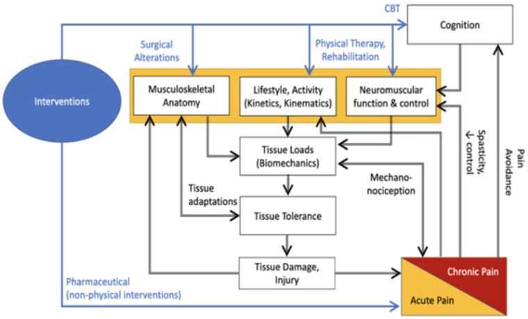
Objective: Biomechanics represents the common final output through which all biopsychosocial constructs of back pain must pass, making it a rich target for phenotyping. To exploit this feature, several sites within the NIH Back Pain Consortium (BACPAC) have developed biomechanics measurement and phenotyping tools. The overall aims of this article were to: 1) provide a narrative review of biomechanics as a phenotyping tool; 2) describe the diverse array of tools and outcome measures that exist within BACPAC; and 3) highlight how leveraging these technologies with the other data collected within BACPAC could elucidate the relationship between biomechanics and other metrics used to characterize low back pain (LBP).

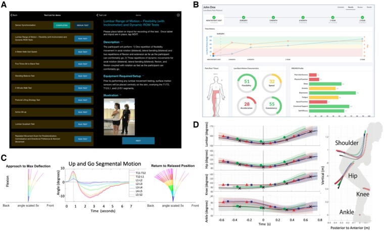
Methods: The narrative review highlights how biomechanical outcomes can discriminate between those with and without LBP, as well as among levels of severity of LBP. It also addresses how biomechanical outcomes track with functional improvements in LBP. Additionally, we present the clinical use case for biomechanical outcome measures that can be met via emerging technologies.

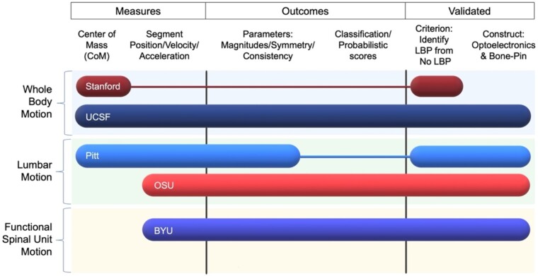
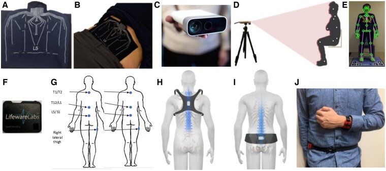
Results: To answer the need for measuring biomechanical performance, our "Results" section describes the spectrum of technologies that have been developed and are being used within BACPAC.

Conclusion and future directions: The outcome measures collected by these technologies will be an integral part of longitudinal and cross-sectional studies conducted in BACPAC. Linking these measures with other biopsychosocial data collected within BACPAC increases our potential to use biomechanics as a tool for understanding the mechanisms of LBP, phenotyping unique LBP subgroups, and matching these individuals with an appropriate treatment paradigm.

Keywords: Biomechanics; Human Movement; Low Back Disorders; Low Back Pain; Motion Analysis Technology.


Figures

Figure 1.
Figure 1.
BACPAC Biomechanics Working Group theoretical model of the role of biomechanics in low back pain. This framework posits that the biopsychosocial elements of LBP (in orange) directly and indirectly (via cognition) influence and are influenced by tissue loading during static or dynamic tasks, thereby changing how an individual moves. Knowledge of these interrelationships can assist in proposing targeted interventions (shown in blue).
Figure 2.
Figure 2.
Basic overview and comparison of technologies used by BACPAC sites. In this figure, rows indicate the tool used by each site and the tool's general spatial complexity. Three factors were considered for columns. Measures determined whether the tool acts regionally or describes whole-body motion. Outcomes provide a general overview of the anticipated process and deliver outcome measures to a patient or clinician. Validation provides whether the tool has successfully demonstrated suitable criterion and construct validity.
Figure 3.
Figure 3.
Visual of the technology to measure biomechanical performance included in BACPAC. Plot (A) depicts the SPINE Sense System developed by Brigham Young University that (B) is adhered onto a participant's skin to track localized lumbar skin strain fields, which correlate with underlying motion of lumbar functional spinal units. The University of California, San Francisco, has leveraged markerless-based kinect cameras (C) to estimate joint centers, thus capturing whole-body sagittal plane motion (D), which can be combined with pressure mats (E) to calculate whole-body kinetics. The University of Pittsburgh adheres off-the-shelf Lifeware IMUs (F) to the skin over the right thigh and various spinous processes (G) to measure both hip and lumbopelvic kinematics. Ohio State University has miniaturized their existing lumbar motion monitor mounting accelerometers to a chest-mounted harness (H) and pelvis belt (I) to quantify lumbopelvic motion with a system that can be worn over the clothes. Using accelerometers to characterize general activity levels, Stanford University uses a dual-mounted accelerometer system (J) to determine the best accelerometer placement and activity thresholding to improve the phenotyping capabilities of existing actigraphy.
Figure 4.
Figure 4.
Example of system interfaces. Many system interfaces, such as the LB3P developed by University of Pittsburgh, provide an interface to clinicians to select specific tasks (top left). The interface provides a brief task description that can be expanded to include specific instructions to coach participants and therapists through a series of specific exercises (A). Furthermore, many of these systems immediately display measures to the clinician and participant in real time. The interface from Ohio State University (B) displays discrete measures of low back motion characteristics of a patient and how they change over time. Brigham Young University captures motion at each lumbar spine segment (C), with the capability to break a complex task such as a timed Up and Go into phases to display how vertebral bodies move when a patient returns to a relaxed position. Systems such as that used by the University of California, San Francisco, provide a technology to allow clinicians to evaluate comprehensive temporal complexity at multiple joints (D), which can be compared between different populations.

References

    1. Hoy D, March L, Brooks P, et al. The global burden of low back pain: Estimates from the Global Burden of Disease 2010 study. Ann Rheum Dis 2014;73(6):968–74. - PubMed
    1. Knezevic NN, Candido KD, Vlaeyen JWS, Zundert JV, Cohen SP.. Low back pain. Lancet 2021;398(10294):78–92. - PubMed
    1. da Silva T, Mills K, Brown BT, et al. Risk of recurrence of low back pain: A systematic review. J Orthop Sport Phys 2017;47(5):305–13. - PubMed
    1. Marras WS. The complex spine: The multidimensional system of causal pathways for low-back disorders. Hum Factors J Hum Factors Ergonom Soc 2012;54(6):881–9. - PubMed
    1. James SL, Abate D, Abate KH, et al. Global, regional, and national incidence, prevalence, and years lived with disability for 354 diseases and injuries for 195 countries and territories, 1990–2017: A systematic analysis for the Global Burden of Disease Study 2017. Lancet Lond Engl 2018;392(10159):1789–858. - PMC - PubMed

Publication types