Case Reports
. 2022 Oct 31:23:e937826.
doi: 10.12659/AJCR.937826.

Neck pain and Headache Complicated by Persistent Syringomyelia After Foramen Magnum Decompression for Chiari I Malformation: Improvement with Multimodal Chiropractic Therapies

Affiliations
Case Reports

Neck pain and Headache Complicated by Persistent Syringomyelia After Foramen Magnum Decompression for Chiari I Malformation: Improvement with Multimodal Chiropractic Therapies

Eric Chun-Pu Chu et al. Am J Case Rep. .

Abstract

BACKGROUND Patients with Arnold-Chiari Malformation I (CM-I) treated with foramen magnum decompression (FMD) can have ongoing neck pain, headaches, and other symptoms complicated by persistent syringomyelia, yet there is little research regarding treatment of these symptoms. CASE REPORT A 62-year-old woman with a history of residual syringomyelia following FMD and ventriculoperitoneal shunt for CM-I presented to a chiropractor with progressively worsening neck pain, occipital headache, upper extremity numbness and weakness, and gait abnormality, with a World Health Organization Quality of Life score (WHO-QOL) of 52%. Symptoms were improved by FMD 16 years prior, then progressively worsened, and had resisted other forms of treatment, including exercises, acupuncture, and medications. Examination by the chiropractor revealed upper extremity neurologic deficits, including grip strength. The chiropractor ordered whole spine magnetic resonance imaging, which demonstrated a persistent cervico-thoracic syrinx and findings of cervical spondylosis, and treated the patient using a multimodal approach, with gentle cervical spine mobilization, soft tissue manipulation, and core and finger muscle rehabilitative exercises. The patient responded positively, and at the 6-month follow-up her WHO-QOL score was 80%, her grip strength and forward head position had improved, and she was now able to eat using chopsticks. CONCLUSIONS This case highlights a patient with neck pain, headaches, and persistent syringomyelia after FMD for CM-I who improved following multimodal chiropractic and rehabilitative therapies. Given the limited, low-level evidence for these interventions in patients with persistent symptoms and syringomyelia after FMD, these therapies cannot be broadly recommended, yet could be considered on a case-by-case basis.


Conflict of interest statement

Conflict of interest: None declared

Figures

Figure 1.
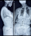
Figure 1.
Full spine radiographs taken at an outside facility prior to presentation to the chiropractor. In the lateral view (A), the patient is noted to have an anterior head position with loss of the cervical lordosis. There is evidence of suboccipital craniectomy and laminectomy of C1 and C2. In the anteroposterior view (B), 3 scoliotic curvatures are evident, including a 20° levoconvex cervicothoracic curvature (upper Θ), a 20° dextroconvex lumbar curvature (lower Θ), and 13° levoconvex thoracic curvature (not shown).The visible span of the ventriculoperitoneal shunt appears intact in both views (arrows).
Figure 2.
Figure 2.
Sagittal cervical spine magnetic resonance imaging. Most evident on the T1-weighted image (A), bone defects are noted around the foramen magnum and posterior elements of C1 and C2 (bracket; }), suggestive of suboccipital craniectomy and laminectomy of C1 and C2. Also evident on the T1-weighted and T2-weighted image (B) is a spinal cord syrinx (arrowheads). At the lowermost extent of the image, the upper thoracic spinal canal is not visible, given the patient’s scoliotic curvature at this region.
Figure 3.
Figure 3.
Axial T2-weighted cervical magnetic resonance image at C4/5. There is moderate narrowing of the central canal due to an eccentric disc osteophyte complex on the right (arrow). There is also moderate narrowing of the right neural foramen due to disc herniation (arrowhead).
Figure 4.
Figure 4.
Right oblique sagittal proton density turbo spin echo cervical magnetic resonance image. Moderate narrowing of the right C4/5 and C6/7 neural foramina due to disc herniation is evident (arrows).
Figure 5.
Figure 5.
Cervical mobilization demonstration. The provider applies gentle posterior to anterior pressure to the left mid-cervical spine while the opposite hand ipsilaterally laterally flexes and slightly rotates the head and stabilizes it against the head rest. This procedure is repeated on the contralateral side as well as in the lower cervical and upper thoracic spine (not shown).
Figure 6.
Figure 6.
Instrument-assisted soft tissue manipulation. The provider applies emollient to the skin surface then gently strokes a massage tool (Strig, Korea) across the skin surface (arrows). This treatment was applied in the region of the upper trapezius (shown), as well as the rhomboids and levator scapulae muscles bilaterally.
Figure 7.
Figure 7.
Core strengthening exercise demonstration. The patient sits in this device (Allcore360°, USA) that rotates slowly at different angles of inclination, while the patient isometrically contracts the core muscles to remain in the neutral, seated posture while holding a large ring in the hands and ball between the knees.
Figure 8.
Figure 8.
Hand strength rehabilitation demonstration. The patient is wearing robotic gloves (Ober, China) and conducts fine motor activities for up to 30 min per day. Also shown is the control console that allows the level of mechanical assistance to be altered. Image from EC.

References

    1. van Dellen JR. Chiari malformation: An unhelpful eponym. World Neurosurgery. 2021;156:1–3. - PubMed
    1. George TM, Higginbotham NH. Defining the signs and symptoms of Chiari malformation type I with and without syringomyelia. Neurol Res. 2011;33:240–46. - PubMed
    1. Vandertop WP. Syringomyelia. Neuropediatrics. Georg Thieme Verlag KG. 2014;45:3–9. - PubMed
    1. Schuster JM, Zhang F, Norvell DC, Hermsmeyer JT. Persistent/recurrent syringomyelia after chiari decompression – natural history and management strategies: A systematic review. Evid Based Spine Care J. 2013;4:116–25. - PMC - PubMed
    1. Soleman J, Bartoli A, Korn A, et al. Treatment failure of syringomyelia associated with Chiari I malformation following foramen magnum decompression: How should we proceed? Neurosurg Rev. 2019;42:705–14. - PubMed

Publication types