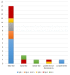
Prevalence of corneal findings and their interrelation with hematological findings in monoclonal gammopathy
- PMID: 36315502
- PMCID: PMC9621422
- DOI: 10.1371/journal.pone.0276048
Prevalence of corneal findings and their interrelation with hematological findings in monoclonal gammopathy
Abstract
Purpose: To determine prevalence of paraproteinemic keratopathy (PPK) among patients with monoclonal gammopathy (MG). To evaluate interrelation between corneal and hematological parameters in patients with PPK.
Methods: Fifty-one patients with monoclonal gammopathy of undetermined significance (n = 19), smoldering multiple myeloma (n = 5) or multiple myeloma (n = 27) were prospectively included in this study. Best-corrected visual acuity, slit-lamp biomicroscopy, Scheimpflug tomography, in-vivo confocal laser scanning microscopy, optical coherence tomography and complete hematological workup were assessed.
Results: We identified n = 19 patients with bilateral corneal opacities compatible with PPK. PPK was newly diagnosed in 13 (29%) of 45 patients with a primary hematological diagnosis and in n = 6 patients without previous hematological diagnosis. The most common form was a discreet stromal flake-like PPK (n = 14 of 19). The median level of M-protein (p = 0.59), IgA (p = 0.53), IgG (p = 0.79) and IgM (p = 0.59) did not differ significantly between the patients with and without PPK. The median level of the FLC κ in serum of patients with kappa-restricted plasma cell dyscrasia was 209 mg/l in patients with PPK compared to 38.1 mg/l in patients without PPK (p = 0.18). Median level of FLC lambda in serum of patients with lambda-restricted plasma cell dyscrasia was lower in patients with PPK compared to patients without PPK (p = 0.02).
Conclusion: The PPK was mostly discreet, but its prevalence (29%) was higher than expected. Median level of the monoclonal paraprotein was not significantly higher in patients with PPK compared to patients without PPK. Our results suggest a lack of correlation between morphology and severity of the ocular findings and severity of the monoclonal gammopathy.
Trial registration: German Clinical Trial Register: DRKS00023893.
Conflict of interest statement
The authors have declared that no competing interests exist.
Figures


References
-
- Blobner F., Kristallinische Degeneration der Bindehaut und Hornhaut. Klin Monatsbl Augenheilkd, 1938. 100: p. 588–593.
-
- Meesmann, Über eine eigenartige Hornhautdegeneration (?) (Ablagerung des Bence-Jonesschen Eiweisskörpers in der Hornhaut), in Bericht über die Fünfzigste Zusammenkunft der Deutschen Ophthalmologischen Gesellschaft in Heidelberg 1934, Wagenmann A., Editor. 1934, Springer Berlin Heidelberg: Berlin, Heidelberg. p. 311–315.
-
- Lisch W., et al.., Chameleon-like appearance of immunotactoid keratopathy. Cornea, 2012. 31(1): p. 55–8. - PubMed
Publication types
MeSH terms
Associated data
LinkOut - more resources
Full Text Sources
Medical
Miscellaneous

