. 2022 Oct 31;17(10):e0269093.
doi: 10.1371/journal.pone.0269093. eCollection 2022.

5-ethyl-2'-deoxyuridine fragilizes Klebsiella pneumoniae outer wall and facilitates intracellular killing by phagocytic cells

Affiliations

5-ethyl-2'-deoxyuridine fragilizes Klebsiella pneumoniae outer wall and facilitates intracellular killing by phagocytic cells

Estelle Ifrid et al. PLoS One. .

Abstract

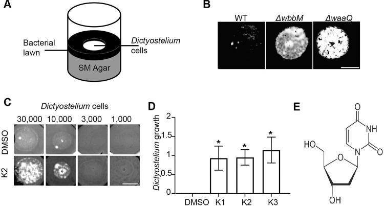
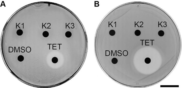
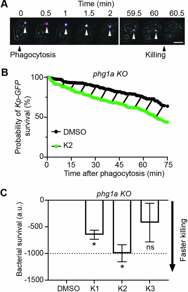
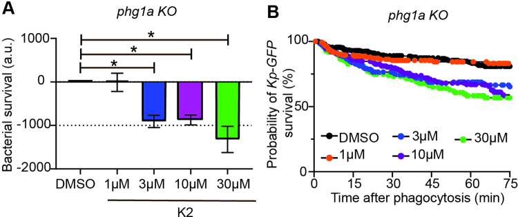
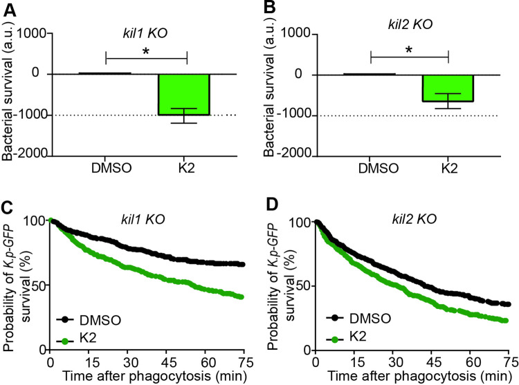
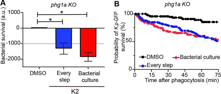
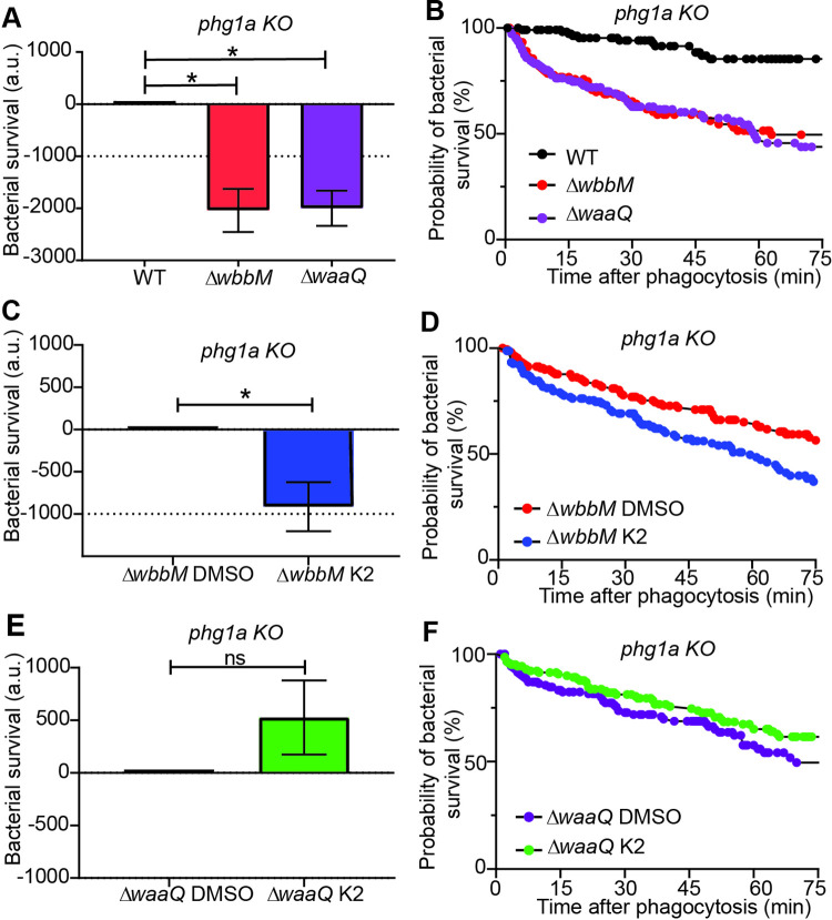
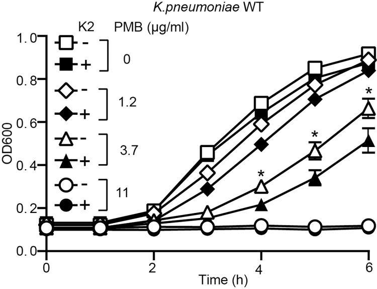
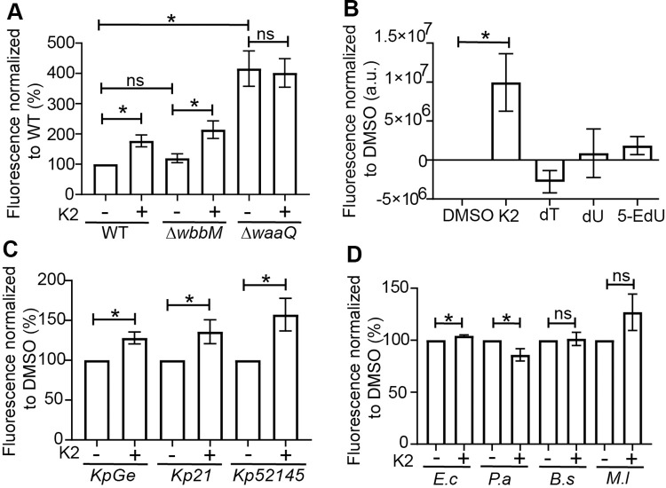
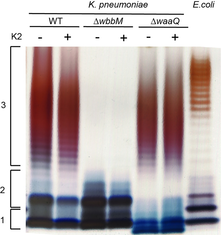
Klebsiella pneumoniae is the causative agent of a variety of severe infections. Many K. pneumoniae strains are resistant to multiple antibiotics, and this situation creates a need for new antibacterial molecules. K. pneumoniae pathogenicity relies largely on its ability to escape phagocytosis and intracellular killing by phagocytic cells. Interfering with these escape mechanisms may allow to decrease bacterial virulence and to combat infections. In this study, we used Dictyostelium discoideum as a model phagocyte to screen a collection of 1,099 chemical compounds. Phg1A KO D. discoideum cells cannot feed upon K. pneumoniae bacteria, unless bacteria bear mutations decreasing their virulence. We identified 3 non-antibiotic compounds that restored growth of phg1A KO cells on K. pneumoniae, and we characterized the mode of action of one of them, 5-ethyl-2'-deoxyuridine (K2). K2-treated bacteria were more rapidly killed in D. discoideum phagosomes than non-treated bacteria. They were more sensitive to polymyxin and their outer membrane was more accessible to a hydrophobic fluorescent probe. These results suggest that K2 acts by rendering the membrane of K. pneumoniae accessible to antibacterial effectors. K2 was effective on three different K. pneumoniae strains, and acted at concentrations as low as 3 μM. K2 has previously been used to treat viral infections but its precise molecular mechanism of action in K. pneumoniae remains to be determined.


Conflict of interest statement

The authors have declared that no competing interests exist.

Figures

Fig 1
Fig 1. Three compounds affect the interaction between K. pneumoniae and phagocytic amoebae.
A. D. discoideum cells were deposited on a lawn of K. pneumoniae and allowed to form a phagocytic plaque for 10 days. B. Phg1A KO cells failed to grow on WT bacteria, but they grew readily on K. pneumoniae mutants with decreased virulence (ΔwaaQ, ΔwbbM) (scale bar: 4mm). C. K2 increased the ability of phg1a KO cells to create phagocytic plaques in comparison with the negative control (DMSO) (scale bar: 4mm). D. The effect of each compound was scored from 4 (visible growth of 1,000 cells) to 0 (no growth of 30,000 cells) and the score of the negative control (DMSO) substracted. In this scale, the result shown in Fig 1C would score as a 0 for DMSO, and 2 for K2. Repeated experiments showed a high variability, but a significant effect for all three selected compounds (mean ± SEM; *: p<0.05; Kruskal-Wallis test, Dunn’s test. DMSO, K2: N = 10; K1, K3: N = 7 independent experiments). The original uncontrasted pictures are shown in S1 Fig in S1 File. E. Chemical structure of the K2 compound.
Fig 2
Fig 2. Selected compounds exhibit no antibiotic activity against K. pneumoniae.
K. pneumoniae were plated on LB- (A) or SM- (B) agar plates. Paper discs with 20 μl of a 10 mM DMSO stock solution of each compound were then placed on the agar and the bacteria allowed to grow. After an overnight incubation at 25°C, a halo of bacterial growth inhibition was observed around a disk containing tetracycline (TET), but none of the selected compounds showed a similar effect (scale bar: 2 cm).
Fig 3
Fig 3. K2 increases the intracellular killing of K. pneumoniae by phg1a KO cells.
A. Time-lapse images showing one representative example of a fluorescent K. pneumoniae ingested by a D. discoideum phg1a KO cell. The phase contrast and fluorescence pictures were superimposed, and the position of the fluorescent bacteria indicated with an arrowhead. Time 0 is defined as the time when the D. discoideum cell engulfs the bacteria (Phagocytosis). In this example, extinction of fluorescence was observed 60 minutes after ingestion (Killing) (scale bar: 6μm). Only the essential time points are shown, showing the moment when the bacteria was phagocytosed (t = 0), and when their fluorescence was lost (t = 60min). B. Survival of K. pneumoniae (%, Kaplan-Meier estimator) ingested by phg1a KO cells was decreased in the presence of the compound K2 (green) compared with the DMSO control (black). These two curves were obtained by combining the results of 6 independent experiments (total 180 bacteria for each condition). The dashed area represents the difference between the two survival curves. C. The Kaplan-Meier survival curves were determined in multiple independent experiments (30 bacteria for each condition) for the three tested compounds (30μM). The area under the curve (AUC) for each compound (K1-K3) and the control (DMSO) was determined for each experiment over 75 min, and the difference (corresponding to the dashed area in Fig 3B) was calculated. A figure inferior to zero indicates that the killing was faster in the presence of the compound than in the control (DMSO) condition. Two compounds, K1 and K2, increased significantly the intracellular killing of K. pneumoniae by phg1a KO cells (mean ± SEM; *: p<0.05; Kruskal-Wallis test, Dunn’s test. K1, K3: N = 3; DMSO; K2: N = 11 independent experiments).
Fig 4
Fig 4. K2 is active at a concentration of 3 μM and above.
A. The effect of K2 on the intracellular killing of K. pneumoniae was determined as described in the legend to Fig 3 at concentrations of K2 ranging from 1 to 30 μM. K2 increased intracellular killing of bacteria at 3 μM (blue), 10 μM (purple) and 30 μM (green) (mean ± SEM; *: p<0.05; Kruskal-Wallis test, Dunn’s test. DMSO, 3 μM and 10 μM: N = 6; 1 μM: N = 5; 30 μM: N = 4 independent experiments). B. The corresponding survival curves of ingested K. pneumoniae are shown.
Fig 5
Fig 5. K2 increases intracellular killing of K. pneumoniae by kil1 KO and kil2 KO cells.
The effect of the K2 compound on K. pneumoniae intracellular killing was assessed after ingestion by kil1 KO cells (A) or by kil2 KO cells (B). K2 (green) increased significantly bacterial killing in both mutant cells (mean ± SEM; * p<0.05 Mann-whitney test, kil1 KO: N = 9; kil2 KO: N = 13 independent experiments). C, D. The corresponding survival curves of ingested K. pneumoniae in kil1 KO (C) and kil2 KO cells (D) are shown.
Fig 6
Fig 6. K2 treatment renders K. pneumoniae more susceptible to intracellular killing.
A. The effect of K2 on intracellular killing was assessed by adding it at different steps of the experimental process. K2 was added either at every step (bacterial overnight culture, D. discoideum overnight culture and during the ingestion and killing of bacteria; blue), or only in the overnight bacterial culture (red), and compared to a treatment with DMSO (mean ± SEM; *: p<0.05; Kruskal-Wallis test, Dunn’s test. N = 5 independent experiments). B. The corresponding survival curves with K2 added at every step (blue) or only during the bacterial preculture (red) are shown.
Fig 7
Fig 7. Intracellular killing of K. pneumoniae mutants.
A. Intracellular killing of K. pneumoniae (WT: black, ΔwbbM: red, ΔwaaQ: purple) in phg1A KO D. discoideum was assessed as described in the legend to Fig 3 (mean ± SEM; *: p < 0.05; Mann-whitney test. ΔwbbM: N = 4; ΔwaaQ; N = 5 independent experiments) B. The corresponding survival curves of ΔwbbM (red), ΔwaaQ (purple) and WT K. pneumoniae (black) are shown. C. Exposure to K2 further increased the intracellular killing of ΔwbbM K.pneumoniae in phg1a KO cells (mean ± SEM; *: p < 0.05; Mann-whitney test. N = 5 independent experiments). D. The corresponding survival curves of ΔwbbM K. pneumoniae treated with DMSO (red) or K2 (blue) are shown. E. Exposure to K2 did not further increase intracellular killing of ΔwaaQ K. pneumoniae in phg1a KO cells (mean ± SEM; Mann-whitney test. N = 5 independent experiments). F. The corresponding survival curves of ΔwaaQ K. pneumoniae treated with DMSO (purple) or K2 (green) are shown.
Fig 8
Fig 8. K2 increases the sensitivity of K. pneumoniae to polymyxin B.
WT K. pneumoniae were grown overnight in the presence or absence of K2. The bacteria were then diluted and their growth was assessed for 6 h in the continued presence or absence of K2 and in the presence of increasing concentrations of polymyxin B (PMB: 0-11 μg/ml). While exposure to K2 did not alter the growth of K. pneumoniae, it increased its sensitivity to polymyxin B as evidenced by a slower growth in the presence of 1.2 or 3.7 μg/ml of PMB (mean ± SEM; *: p < 0.05; Mann-whitney test. PMB 3.7 μg/ml: N = 12 independent experiments).
Fig 9
Fig 9. K2 treatment increases access of a hydrophobic probe to the bacterial membrane of K. pneumoniae.
A. In order to assess the efficacy with which the LPS shielded the bacterial membrane, bacteria were exposed to the fluorescent probe 1-N-phenylnaphthylamine (NPN), and fluorescence was recorded, providing a measure of the insertion of NPN in the bacterial membrane. The membrane of ΔwaaQ mutant K. pneumoniae was more accessible to NPN than that of WT and ΔwbbM mutant. In K2-treated bacteria, access of NPN increased in WT and in ΔwbbM but not in ΔwaaQ bacteria (mean ± SEM; *: p<0.05; Kruskal-Wallis test; N = 5 independent experiments). B. Three chemical analogs of K2 (dT = deoxythymidine, dU = deoxyuridine and 5-EdU = 5-Ethynyl-2’-deoxyuridine) were tested for their ability to increase the membrane accessibility of NPN. K2 was the only compound that increased significantly the accessibility of the outer membrane of K. pneumoniae to NPN (mean ± SEM; *: p<0.05; Kruskal-Wallis test; DMSO, K2: N = 6; and N = 5 independent experiments for the three analogs of K2). C, D. The effect of K2 was tested on three different strains of K. pneumoniae (KpGE, Kp21 and Kp52145) as well as Escherichia coli (E.c), Pseudomonas aeruginosa (P.a), Bacillus subtilis (B.s) and Microccocus luteus (M.l). K2 increased NPN incorporation in K. pneumoniae, but exhibited little or no effect on other bacteria. (mean ± SEM; *: p<0.05; Mann-whitney test. N = 8 independent experiments).
Fig 10
Fig 10. K2 treatment does not visibly affect LPS structure.
Bacteria were grown overnight in the presence or absence of K2. LPS from K. pneumoniae WT and mutant strains (ΔwbbM, ΔwaaQ) were then purified and analyzed by SDS-PAGE electrophoresis and silver staining. LPS extracted from E. coli (serotype O111:B4) was used for comparison. The LPS of WT K. pneumoniae showed the three main forms of the LPS: lipid A (1), lipid A+ oligosaccharide core (2), lipid A + core + O-antigen [27]. As expected, the LPS from ΔwbbM bacteria lacked the O-antigen, and ΔwaaQ displayed a smaller oligosaccharide core that migrated further in the gel. No visible alteration of LPS structure was observed in K2-treated bacteria.

Similar articles

Cited by

References

    1. Agyeman AA, Bergen PJ, Rao GG, Nation RL, Landersdorfer CB. 2019. A systematic review and meta-analysis of treatment outcomes following antibiotic therapy among patients with carbapenem-resistant Klebsiella pneumoniae infections. Int J Antimicrob Agents 55:105833. doi: 10.1016/j.ijantimicag.2019.10.014 - DOI - PubMed
    1. Payne DJ, Gwynn MN, Holmes DJ, Pompliano DL. 2007. Drugs for bad bugs: confronting the challenges of antibacterial discovery. Nat Rev Drug Discov 6:29–40. doi: 10.1038/nrd2201 - DOI - PubMed
    1. Cegelski L, Marshall GR, Eldridge GR, Hultgren SJ. 2008. The biology and future prospects of antivirulence therapies. Nat Rev Microbiol 6:17–27. doi: 10.1038/nrmicro1818 - DOI - PMC - PubMed
    1. Cosson P, Zulianello L, Join-Lambert O, Faurisson F, Gebbie L, Benghezal M,. 2002. Pseudomonas aeruginosa virulence analyzed in a Dictyostelium discoideum host system. J Bacteriol 184:3027–33. doi: 10.1128/JB.184.11.3027-3033.2002 - DOI - PMC - PubMed
    1. Swart AL, Harrison CF, Eichinger L, Steinert M, Hilbi H. 2018. Acanthamoeba and Dictyostelium as Cellular Models for Legionella Infection. Front Cell Infect Microbiol 8:61. doi: 10.3389/fcimb.2018.00061 - DOI - PMC - PubMed

Publication types