. 2023 Jan 12;141(2):194-199.
doi: 10.1182/blood.2022016218.

Dietary iron restriction protects against vaso-occlusion and organ damage in murine sickle cell disease

Affiliations

Dietary iron restriction protects against vaso-occlusion and organ damage in murine sickle cell disease

Huihui Li et al. Blood. .

Abstract

Sickle cell disease (SCD) is an inherited disorder resulting from a β-globin gene mutation, and SCD patients experience erythrocyte sickling, vaso-occlusive episodes (VOE), and progressive organ damage. Chronic hemolysis, inflammation, and repeated red blood cell transfusions in SCD can disrupt iron homeostasis. Patients who receive multiple blood transfusions develop iron overload, and another subpopulation of SCD patients manifest iron deficiency. To elucidate connections between dietary iron, the microbiome, and SCD pathogenesis, we treated SCD mice with an iron-restricted diet (IRD). IRD treatment reduced iron availability and hemolysis, decreased acute VOE, and ameliorated chronic organ damage in SCD mice. Our results extend previous studies indicating that the gut microbiota regulate disease in SCD mice. IRD alters microbiota load and improves gut integrity, together preventing crosstalk between the gut microbiome and inflammatory factors such as aged neutrophils, dampening VOE, and organ damage. These findings provide strong evidence for the therapeutic potential of manipulating iron homeostasis and the gut microbiome to ameliorate SCD pathophysiology. Many treatments, which are under development, focus on lowering the systemic iron concentration to relieve disease complications, and our data suggest that iron-induced changes in microbiota load and gut integrity are related- and novel-therapeutic targets.


Conflict of interest statement

Conflict-of-interest disclosure: The authors declare no relevant competing financial interests.

Figures

None
Graphical abstract
Figure 1.
Figure 1.
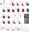
Dietary ironrestriction amelioratesacute VOE in SCD mice. (A) Schematic experimental design for iron diet treatments. (B) Non-heme iron content of the bone marrow (N = 5), spleen (N = 5), and liver (N = 5), and serum iron concentration (N = 10) are decreased after dietary iron restriction. (C) WBC count, (D) RBC count, and total hemoglobin (HGB) concentration in Ctrl and IRD SCD mice (N = 14-15). (E) Mean corpuscular hemoglobin concentration (MCHC), MCH, mean corpuscular volume in Ctrl and IRD mice (merged data from 3 experiments, N = 14-15). (F) Serum soluble adhesion molecules (sP-selectin, sE-selectin, sVcam-1 and sIcam-1) by enzyme-linked immunosorbent assay (N = 10). (G) FACS plots with gating strategy and quantification of CXCR4Hi CD62Lo aged neutrophils in Ctrl and IRD mice. (H) Intravital microscopy images of inflamed cremasteric venules in Ctrl and IRD mice. Scale bars, 10 μm. (I) Cremasteric venule diameters, (J) red cell velocity (Vrbc), (K) blood flow rate, (L) the number of adherent leukocytes (N = 41-47), and (M) survival rate after TNF-α injection (N = 9) are compared between Ctrl and IRD mice. ∗P < .05, ∗∗P < .01, ∗∗∗P < .001.
Figure 2.
Figure 2.
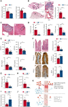
Organ damage parameters are improved in SCD mice after dietary iron restriction. (A) Gross spleen and liver weights are reduced in IRD group compared with Ctrl group. (N = 5) (B) Leukocyte infiltration in lung tissue, quantified by H&E staining, is decreased in IRD treated SCD mice. Low-magnification scale bar (left), 500 μm; high-magnification scale bar (right), 50 μm. (N = 5) (C) H&E staining of liver tissue; area within dashed line shows leukocyte infiltration and necrosis. Scale bar, 100 μm. (D) Liver necrosis, leukocyte infiltration, and fibrosis are all reduced in IRD mice. (N = 4-5) (E) Liver enzymes (alanine aminotransferase and aspartate aminotransferase), total bilirubin concentration, and direct bilirubin concentration from sera of Ctrl and IRD mice. (N = 10-15) (F) Left: representative images of duodenal villi with either H&E, anti-claudin 3 or anti-junctional adhesion molecule A (JAM-A) immunohistology staining in Ctrl and IRD treated SCD mice. H&E scale bar, 50 μm; immunohistology scale bar 20 μm. Right: quantification of intestinal inflammation or percentage of positive stained area. (N = 5) (G) Concentration of FITC-Dextran in serum of IRD mice is significantly lower than Ctrl mice. (N = 7) Dashed line represents levels in the healthy SA mice. (H) Bacterial load measured by Pan-bac quantitative reverse transcription polymerase chain reaction, in fecal samples normalized by the weight of fecal samples. (N = 4-5) Dashed line represents levels in the healthy SA mice. (I) Bacterial Firmicutes/Bacteroidetes ratio of fecal samples from Ctrl and IRD treated SCD mice. (N = 4-5) Dashed line represents levels in the healthy SA mice. (J) Quantification of sera TLR2 ligands activity in Ctrl and IRD treated SCD mice. (N = 5) Dashed line represents levels in the healthy SA mice. (K) Working model. Top: Excess dietary iron expands the proliferation of bacteria (red arrow indicates direct impact from iron) and increases gut permeability (red arrow) which allows the translocation of microbial compounds to internal organs (dashed arrow indicates indirect impact from iron); in parallel, excess iron is absorbed and deposited into organs (red arrow); together lead to exacerbation of neutrophil aging, hemolysis, vaso-occlusion, and organ damage. Bottom: dietary iron restriction reduces bacterial proliferation and ameliorates gut permeability, and consequently decreasing the translocation of microbial compounds and limiting systemic iron levels. Collectively, neutrophil aging, hemolysis, vaso-occlusion, and organ damage are found at lower rates. ∗P < .05, ∗∗P < .01, ∗∗∗P <.001.

Comment in

  • (De)ironing out sickle cell disease.
    Das NK, Shah YM. Das NK, et al. Blood. 2023 Jan 12;141(2):129-130. doi: 10.1182/blood.2022018791. Blood. 2023. PMID: 36633882 Free PMC article. No abstract available.

References

    1. Piel FB, Hay SI, Gupta S, Weatherall DJ, Williams TN. Global burden of sickle cell anaemia in children under five, 2010-2050: modelling based on demographics, excess mortality, and interventions. PLoS Med. 2013;10(7) - PMC - PubMed
    1. van Beers EJ, Yang Y, Raghavachari N, et al. Iron, inflammation, and early death in adults with sickle cell disease. Circ Res. 2015;116(2):298–306. - PMC - PubMed
    1. Ballas SK. Iron overload is a determinant of morbidity and mortality in adult patients with sickle cell disease. Semin Hematol. 2001;38(1 Suppl 1):30–36. - PubMed
    1. Koduri PR. Iron in sickle cell disease: a review why less is better. Am J Hematol. 2003;73(1):59–63. - PubMed
    1. Castro O, Haddy TB. Improved survival of iron-deficient patients with sickle erythrocytes. N Engl J Med. 1983;308(9):527. - PubMed

Publication types