. 2022 Dec;7(12):1956-1966.
doi: 10.1038/s41564-022-01243-4. Epub 2022 Oct 31.

Bacteriophage genome engineering with CRISPR-Cas13a

Affiliations

Bacteriophage genome engineering with CRISPR-Cas13a

Jingwen Guan et al. Nat Microbiol. 2022 Dec.

Abstract

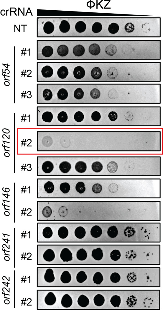
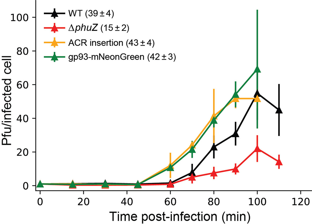
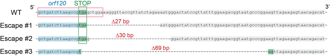
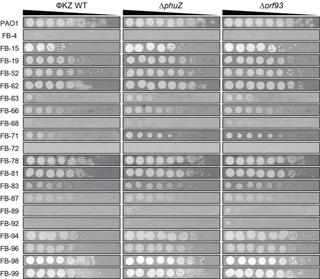
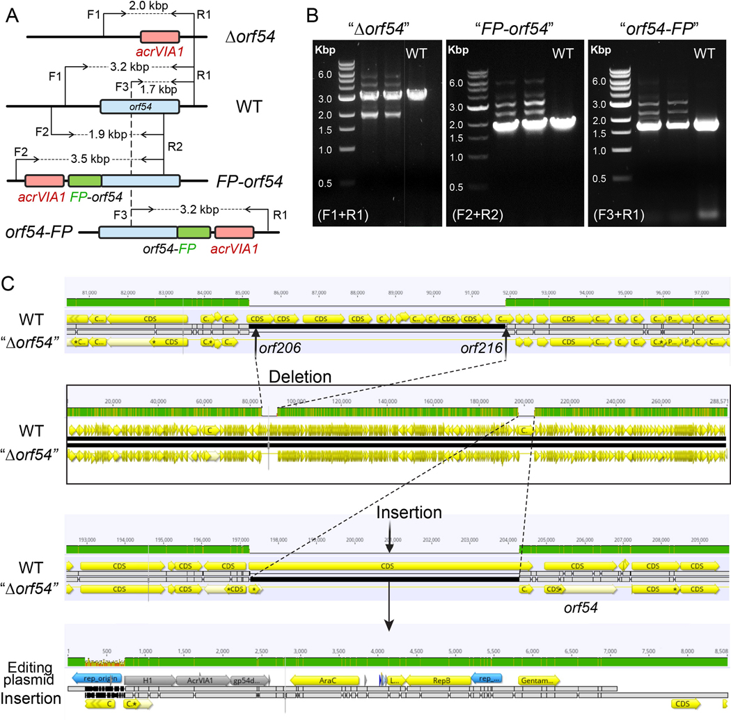
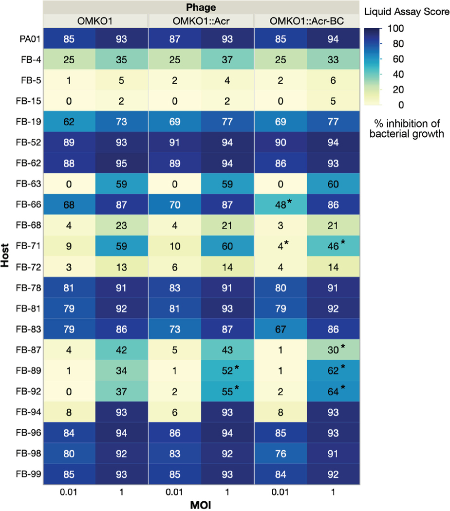
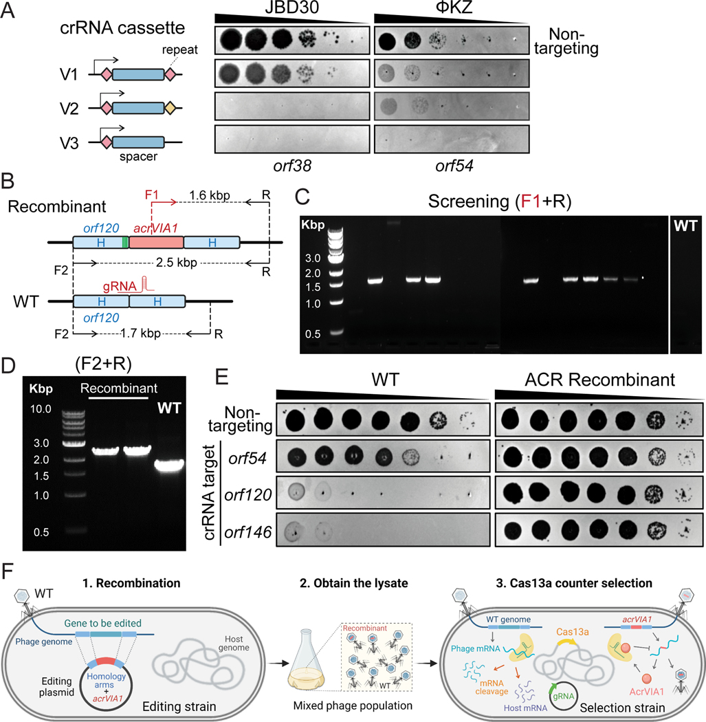
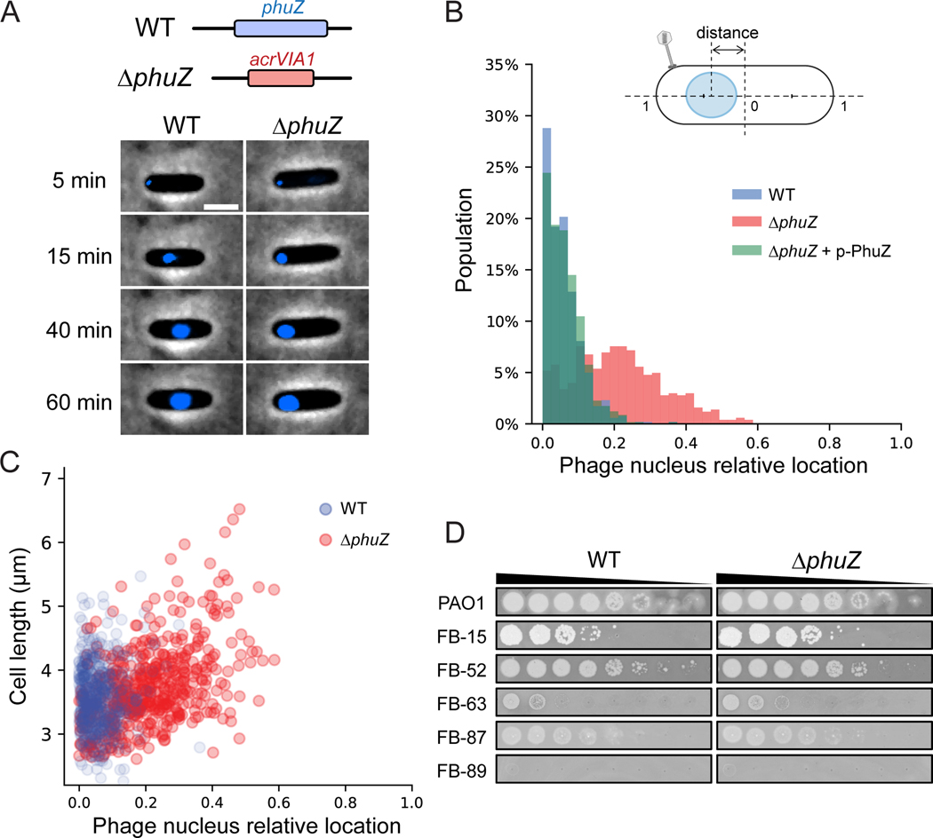
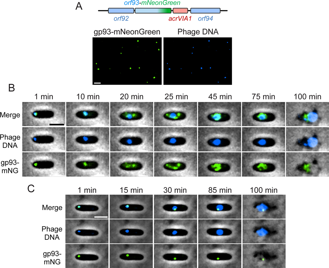
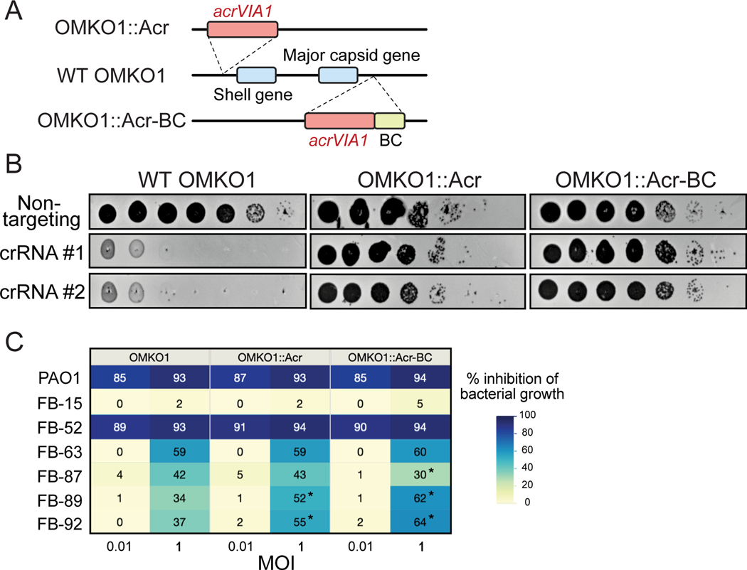
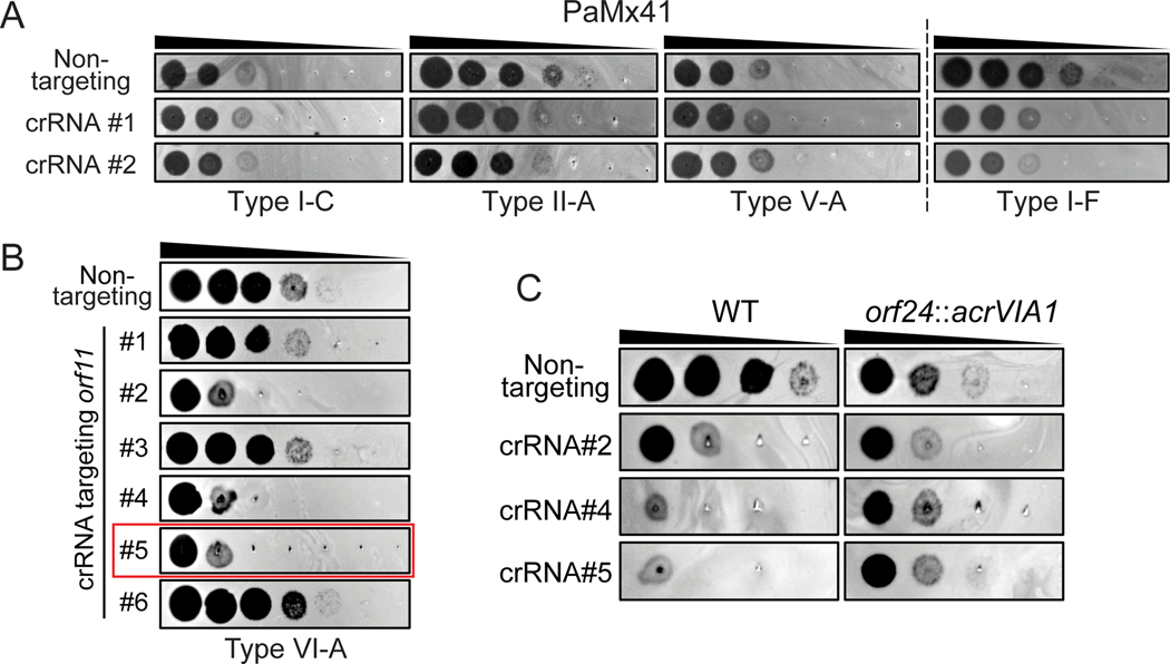
Jumbo phages such as Pseudomonas aeruginosa ФKZ have potential as antimicrobials and as a model for uncovering basic phage biology. Both pursuits are currently limited by a lack of genetic engineering tools due to a proteinaceous 'phage nucleus' structure that protects from DNA-targeting CRISPR-Cas tools. To provide reverse-genetics tools for DNA jumbo phages from this family, we combined homologous recombination with an RNA-targeting CRISPR-Cas13a enzyme and used an anti-CRISPR gene (acrVIA1) as a selectable marker. We showed that this process can insert foreign genes, delete genes and add fluorescent tags to genes in the ФKZ genome. Fluorescent tagging of endogenous gp93 revealed that it is ejected with the phage DNA while deletion of the tubulin-like protein PhuZ surprisingly had only a modest impact on phage burst size. Editing of two other phages that resist DNA-targeting CRISPR-Cas systems was also achieved. RNA-targeting Cas13a holds great promise for becoming a universal genetic editing tool for intractable phages, enabling the systematic study of phage genes of unknown function.


Figures

Extended Data Fig. 1
Extended Data Fig. 1. Plaque efficiency assays of distinct crRNAs of CRISPR-Cas13a targeting transcripts of diverse ΦKZ genes.
Different crRNAs show varied degrees of Cas13a interference efficiency against ΦKZ. The crRNA targeting orf120 highlighted in the red frame has been used for ΦKZ genome engineering. NT, non-targeting.
Extended Data Fig. 2
Extended Data Fig. 2. One-step growth curves of engineered ФKZ variants.
One-step growth curve experiment was performed to determine the latent time period and burst size of engineered phages. Representative plots are shown for each phage strain. The burst sizes are shown in brackets after of each ФKZ variant, representing the mean ± standard error of three or four biologically independent replicates.
Extended Data Fig. 3
Extended Data Fig. 3. Sequence alignment of wild type ΦKZ and three escape mutants at the engineered genomic site.
Escape mutants were isolated and verified by PCR and sequencing. The WT orf120 sequence is highlighted in blue and the downstream region is highlighted in grey. The stop codon (TGA) of orf120 is highlighted in green and Escape mutant #3 reconstitutes it to TAG. The sequence in the red frame matches the spacer sequence of the crRNA that was used to target and eliminate WT phages. Deletions were indicated by dashed lines and their corresponding numbers of absent base pairs.
Extended Data Fig. 4
Extended Data Fig. 4. Determination of host range of ΦKZ ΔphuZ and Δorf93 mutants by plaque assay on P. aeruginosa clinical strains.
Spot-titration of the indicated ΦKZ phages on lawns of clinical isolates of P. aeruginosa (FB-XX).
Extended Data Fig. 5
Extended Data Fig. 5. Failure of genetic editing the shell gene (orf54) in ΦKZ.
(A) Schematic of genomes of WT ΦKZ and three mutated orf54 variants, “Δorf54”, “FP-orf54”, and “orf54-FP”, at the editing site. orf54, acrVIA1, and fluorescent protein (FP) are shown as blue, red, and green rectangles, respectively. F and R indicate forward and reverse primers, respectively, for PCR confirmation of orf54 engineering. (B) PCR confirmation of the indicated orf54 mutants using their corresponding pair of primers. All three mutants generated multiple bands, including a band in the same size as the single band produced by WT. PCR-based screening for engineered ΦKZ orf54 variants have been independently repeated at least three times yielding similar results. (C) Genome alignment of WT phage with the isolated orf54pseudo knock-out” mutant (“Δorf54”). A gene cluster of ~ 7 kbp (orf206 - orf216) was missing in the mutant, likely as a result of phage packaging capacity. The majority of the editing plasmid used to generate recombinants was at the editing site, leaving orf54 intact.
Extended Data Fig. 6
Extended Data Fig. 6. Host range assay of engineered OMKO1 variants.
Host ranges were determined by microplate liquid assay at MOI of 0.01 and 1 on 22 P. aeruginosa clinical strains. The values are presented as the mean liquid assay scores across three independent experiments. Asterisks (*) indicate significant difference between WT and engineered strains as determined by two-sided Students’ T-tests (p < 0.05). The color intensity of each phage-host combination reflects the liquid assay score, which represents how well the phage strain can repress the growth of a given bacterial host. No inhibition of bacterial growth is reflected by a liquid assay score of 0, and complete suppression would result in a score of 100.
Fig. 1:
Fig. 1:. Screening for ΦKZ recombinants by CRISPR-Cas13a counter selection.
(A) Schematic of three versions of CRISPR-Cas13a crRNA cassette and their efficiency of plating assays targeting two unrelated phage strains: JBD30 and ΦKZ. Spacers are represented by dark blue rectangles. Repeats are shown as diamonds with different colors (pink and yellow) indicating different repeat sequences. Arrows mean transcription start sites of PBAD promoter. The cassettes carried the same spacer sequences targeting transcripts of orf38 of JBD30 and orf54 of ΦKZ, respectively. (B) Schematic of WT ΦKZ and recombinant genomes at the editing site. The acrVIA1 gene, shown as a red rectangle, was inserted downstream of orf120, with up-and downstream of homology arms (H) indicated by light blue rectangles. Green stripes represent synonymous mutations that were introduced to the homology region in only this case. Synonymous mutations were not necessary due to the effectiveness of the AcrVIA1 marker on inhibition of Cas13a activities. F and R indicate forward and reverse primers, respectively, being used to confirm the insertion of acrVIA1. (C) Recombinant phages were screened by PCR using primer F1 and R. 8 out of the 16 tested phage plaques generated the expected 1.6 kbp band, which was not detectable in a WT plaque. (D) Recombinant phage plaques underwent 3 rounds of purification and were further confirmed by PCR using primer F2 and R. The PCR products revealed the expected size increase, from ~1.7 kbp of WT to ~2.5 kbp of recombinants (E) Plaque assays showing robust anti-CRISPR activities acquired by recombinant ΦKZ against distinct crRNAs, owing to the successful expression and execution of the incorporated AcrVIA1. PCR-based mutant screening, as well as the subsequent plaque assays, have been independently repeated three times yielding similar results. (F) Workflow of phage genome engineering using CRISPR-Cas13a. 1. an editing plasmid introduces desired genetic modifications via recombination, 2. a mixed phage lysate is generated, 3. the lysate is plated on the selection strain harboring Cas13a and a crRNA targeting WT phages.
Fig. 2:
Fig. 2:. Absence of PhuZ causes mispositioning of the phage nucleus.
(A) Top: schematic of ΦKZ WT and phuZ::acrVIA1phuZ) mutant genomes at the editing site. Bottom: Representative cells infected by WT (left) and ΔphuZ mutant (right). Phage DNA is stained by DAPI and shown as blue signals. The phage nucleus is mispositioned near the cell polar region upon infection by ΔphuZ, in contrast to the WT infection where the phage nucleus is centered. Scale bar denotes 2 μm. (B) Distribution of subcellular location of the phage nucleus in PAO1 cells infected by WT (blue, N = 521) and ΔphuZ (red, N = 503), and a PAO1 strain expressing wild-type PhuZ in trans (p-PhuZ) and infected by ΔphuZ (light green, N = 573). The diagram of an infected cell is shown on the top. The phage nucleus location is defined as the relative distance between the cell center and the nucleus center. (C) The phage nucleus location is plotted against the cell length for WT and ΔphuZ. The phage nucleus position in ΔphuZ-infected cells positively correlates with the cell length with a Pearson correlation coefficient of 0.486, p < 0.001 (two-sided), whereas there is no such correlation for WT infection with a Pearson correlation coefficient of 0.029, p = 0.505 (two-sided). (D) Efficiency of plating of WT and ΔphuZ on representative P. aeruginosa clinical strains. (The full panel of plaque assays is presented in Extended Data Fig. 4)
Fig. 3:
Fig. 3:. Fluorescent labeling of an inner body protein of ΦKZ.
(A) Top: schematic of ΦKZ orf93-mNeonGreen mutant at the editing site. The acrVIA1 gene was inserted downstream of the orf93-mNeonGreen fusion cassette. Bottom: visualization of individual phage particles under the fluorescence microscope. Each mutant phage particle is visible as a green focus (left), due to the packaging of gp93-mNeonGreen in the capsid. ΦKZ genomic DNA is stained by DAPI (right). mNeonGreen and DAPI signals colocalize very well and individual virions are easily distinguishable. Only ~1.4% of the fluorescent phage particles examined (6 out of 438) lacked the mNeonGreen signal. (B) Overlay images (phase-contrast and fluorescent channels) from a time-lapse movie depicting a representative PAO1 cell being infected by a gp93-mNeonGreen mutant phage. gp93-mNeonGreen and phage DNA are shown as green and blue signals, respectively. The representative cell was chosen out of 1093 infected cells from two independent infection experiments. (C) Overlay images from a time-lapse movie showing a PAO1 cell being infected by a WT ΦKZ loaded with gp93-mNeonGreen fusions. Packaged gp93-mNeonGreen appears as a green focus and remains bound to phage DNA throughout the infection cycle. The representative cell was chosen out of 1045 infected cells from two independent infection experiments. Scale bar denotes 2 μm.
Fig. 4:
Fig. 4:. Genetic engineering of therapeutic jumbo phage OMKO1.
(A) Schematic of genomes of two engineered OMKO1 variants where indicated gene fragments are integrated into the WT genome. OMKO1::Acr, acrVIA1 is inserted upstream of the shell gene. OMKO1::Acr-BC, acrVIA1 is inserted together with a barcode sequence (BC) downstream of the major capsid gene. (B) Plaque assays of WT OMKO1 and mutants. Both engineered variants exhibit robust resistance against distinct CRISPR-Cas13a crRNAs, suggesting that the incorporated acrVIA1 is successfully functioning. crRNA #1 and #2 target OMKO1 homologs of ФKZ orf120 and orf146 transcripts, respectively. (C) Determination of host range of WT OMKO1 and mutants on representative P. aeruginosa clinical strains by microplate liquid assay at MOI of 0.01 and 1. Data are presented as the mean liquid assay scores across three independent experiments. Asterisks (*) indicate significant difference between WT and mutants as determined by two-sided Students’ T-tests (p < 0.05). The color intensity of each phage-host combination reflects the liquid assay score, with the darker color the stronger intensity displaying a greater score. Liquid assay score represents how well the phage strain can repress the growth of a given bacterial host. No inhibition of bacterial growth is reflected by a liquid assay score of 0, while complete suppression would result in a score of 100. (The full table of plaque assays is shown in Extended Data Fig. 6)
Fig. 5:
Fig. 5:. Genetic engineering of Podophage PaMx41.
(A) Plaque assays showing PaMx41 resisting a broad variety of DNA-targeting CRISPR-Cas systems, while being (B) targeted by CRISPR-Cas13a. The crRNA sequences of different CRISPR-Cas systems were listed in Table S3. crRNA#5 highlighted in the red frame has been used for PaMx41 genome engineering. (C) PaMx41 orf24::acrVIA1 mutant strain is resistant to diverse crRNAs of CRISPR-Cas13a, due to the expression of AcrVIA1 from the mutant genome. The mutant phages propagated better than WT on the cell lawns of all three crRNAs, though they appeared to exhibit slightly reduced EOP.

References

    1. Kortright KE, Chan BK, Koff JL, and Turner PE (2019). Phage Therapy: A Renewed Approach to Combat Antibiotic-Resistant Bacteria. Cell Host Microbe 25, 219–232. - PubMed
    1. Pires DP, Cleto S, Sillankorva S, Azeredo J, and Lu TK (2016). Genetically Engineered Phages: a Review of Advances over the Last Decade. Microbiol Mol Biol Rev 80, 523–543. - PMC - PubMed
    1. Doss J, Culbertson K, Hahn D, Camacho J, and Barekzi N (2017). A Review of Phage Therapy against Bacterial Pathogens of Aquatic and Terrestrial Organisms. Viruses 9. - PMC - PubMed
    1. Nobrega FL, Costa AR, Kluskens LD, and Azeredo J (2015). Revisiting phage therapy: new applications for old resources. Trends Microbiol 23, 185–191. - PubMed
    1. Lusiak-Szelachowska M, Zaczek M, Weber-Dabrowska B, Miedzybrodzki R, Klak M, Fortuna W, Letkiewicz S, Rogoz P, Szufnarowski K, Jonczyk-Matysiak E, et al. (2014). Phage Neutralization by Sera of Patients Receiving Phage Therapy. Viral Immunol 27, 295–304. - PMC - PubMed

Publication types