. 2022 Nov 1;6(1):24.
doi: 10.1186/s41824-022-00144-3.

PET/MR: primary inferior vena cava leiomyosarcoma

Affiliations

PET/MR: primary inferior vena cava leiomyosarcoma

Brunela Ronchi et al. Eur J Hybrid Imaging. .

Abstract

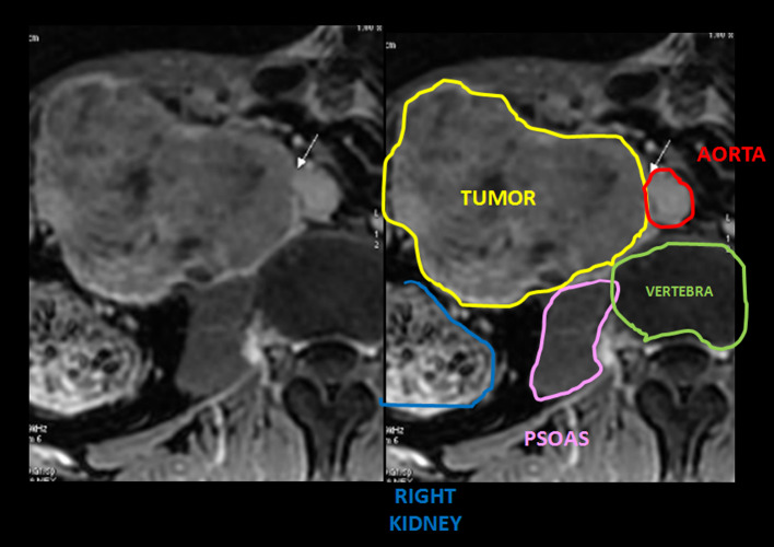
Positron emission tomography (PET) combined with a magnetic resonance (MR) scanner (PET/MR) with 18F-fluorodeoxyglucose (FDG) tracer is being used in quite a few nuclear medicine centers. The aim of this study is to illustrate two uncommon cases of primary inferior vena cava leiomyosarcoma which were formerly evaluated with anatomical images such as computed tomography and ultrasound. These techniques were inferior in the definition of the tumor and its characteristics. F-18 FDG PET/MR was essential and provided all the necessary information: its origin, local extension, anatomo-metabolic behavior, form of presentation, and distant metastasis in one single diagnostic technique. PET/MR accurately contributed to the diagnosis in a shortened period of time and, therefore, in the prognosis of this disease with greater benefits.

Keywords: FDG; Leiomyosarcoma; PET/MR; Retroperitoneal tumor; Vascular tumor; Vena cava.


Conflict of interest statement

The authors declare that they have no competing interests.

Figures

Fig. 1
Fig. 1
PET/MRI (A) images illustrate solid and hypermetabolic (elevated FDG uptake SUVmax 11.91) expansive formation in the retroperitoneum. It can be seen as iso-/hypointense on T1 (B) and heterointense on T2 and T2 FAT SAT (C, D respectively), with well-defined and lobulated borders. It presents a heterogeneous enhancement after IV contrast (G), due to necrosis areas, high restriction on diffusion images (EF) and contrast enhancement intensifies in delayed images (H). Negative peak sign from psoas muscle was useful in the diagnosis (see description below) (BD)
Fig. 2
Fig. 2
MIP images A showed a hypermetabolic (black arrow: SUV max 13.95) voluminous expansive mass in the retroperitoneum and abdominal cavity. PET/MRI sagittal fusion images B illustrate the extension and compromise of distant organs. PET/MRI axial images C, D demonstrate the origin of the tumor from IVC (white arrow) with an eccentric growth
Fig. 3
Fig. 3
Mingoli’s anatomical classification of IVC LMS
Fig. 4
Fig. 4
MR axial T1 enhancement scan and schematic representation shows the positive embedded sign seen in our second patient which compresses and displaces neighbor organs like right kidney and liver. This finding was key to locating the origin of the tumor
Fig. 5
Fig. 5
MR axial T2 scan from the first patient and schematic representation illustrates the negative peak sign from psoas muscle, dismissing its origin

Similar articles

Cited by

References

    1. Bednarova I, Frellesen C, Roman A, Vogl TJ. Case 257: leiomyosarcoma of the inferior vena cava. Radiology. 2018;288(3):901–908. doi: 10.1148/radiol.2018160821. - DOI - PubMed
    1. Blum U, Wildanger G, Windfuhr M, Laubenberger J, Freudenberg N, Munzar T. Preoperative CT and MR imaging of inferior vena cava leiomyosarcoma. Eur J Radiol. 1995;20(1):23–27. doi: 10.1016/0720-048x(95)00608-s. - DOI - PubMed
    1. Coindre JM. Grading of soft tissue sarcomas: review and update. Arch Pathol Lab Med. 2006;130(10):1448–1453. doi: 10.5858/2006-130-1448-GOSTSR. - DOI - PubMed
    1. Ghose J, Bhamre R, Mehta N, Desouza A, Patkar S, Dhareshwar J, Goel M, Shrikhande SV. Resection of the inferior vena cava for retroperitoneal sarcoma: six cases and a review of literature. Indian J Surg Oncol. 2018;9(4):538–546. doi: 10.1007/s13193-018-0796-9. - DOI - PMC - PubMed
    1. Molina M, Schiappacasse G, Labra A. Tumors that invade the inferior vena cava: an illustrative review of the main imaging features on computed tomography and magnetic resonance. Rev Chil Radiol. 2016;22(1):39–46.

LinkOut - more resources