

Posterior ocular manifestations following BNT162b2 mRNA COVID-19 vaccine: a case series
- PMID: 36316618
- PMCID: PMC9628305
- DOI: 10.1007/s10792-022-02565-2
Posterior ocular manifestations following BNT162b2 mRNA COVID-19 vaccine: a case series
Abstract
Purpose: To report the occurrence of posterior ocular adverse events following the administration of the BNT162b2 mRNA vaccine against SARS-CoV-2.
Methods: A retrospective consecutive case series, in which the medical files of patients presenting with ocular adverse events within 30 days of the vaccine inoculation, were analyzed.
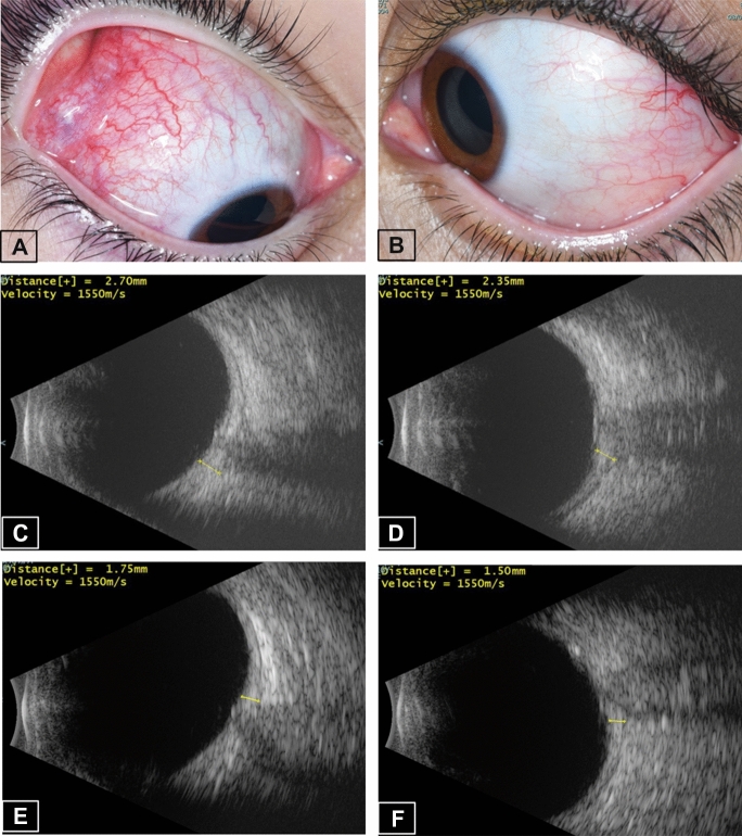
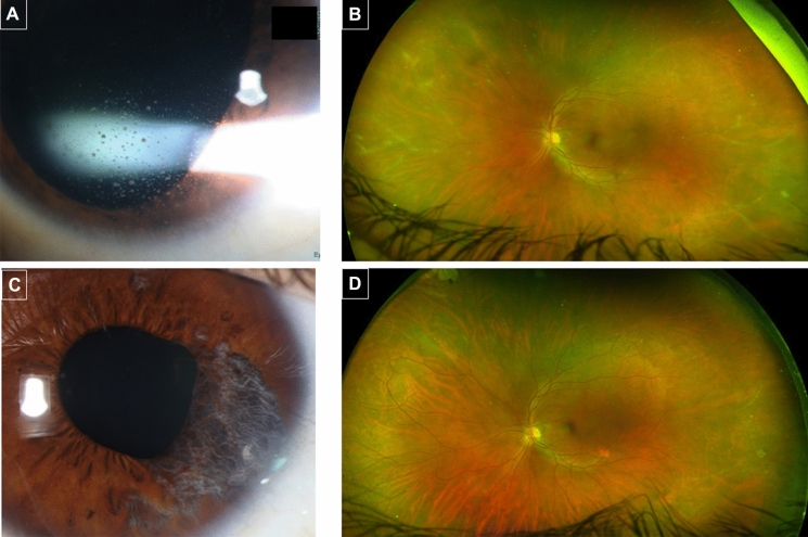
Results: Four patients (2 females) were included in the study. The diagnoses included: posterior scleritis, paracentral acute middle maculopathy, herpes panuveitis, and Vogt-Koyanagi-Harada (VKH)-like uveitis. Three of the patients had no relevant ocular history, but the patient who developed scleritis was in remission without medical therapy for four years, until the flare-up, which occurred one day after the vaccine. All patients improved with treatment.
Conclusion: Though a causal relationship cannot be definitively established, the temporal relationship suggests a possible link between the COVID-19 vaccine and the posterior ocular complications. The benefits of vaccination clearly outweigh the potential adverse effects; however, ophthalmologists should be aware of the potential for vaccine-associated uveitis.
Keywords: COVID-19; Herpes; Ocular inflammation; PAMM; SARS-CoV-2; Scleritis; Uveitis; Vaccine.
© 2022. The Author(s), under exclusive licence to Springer Nature B.V.
Conflict of interest statement
The authors declare no competing interests.
The authors have no relevant financial or non-financial interests to disclose.
Figures


Similar articles
-
Case report: Bilateral panuveitis resembling Vogt-Koyanagi-Harada disease after second dose of BNT162b2 mRNA COVID-19 vaccine.Front Immunol. 2022 Sep 29;13:967972. doi: 10.3389/fimmu.2022.967972. eCollection 2022. Front Immunol. 2022. PMID: 36248859 Free PMC article.
-
Vaccine-Associated Uveitis after COVID-19 Vaccination: Vaccine Adverse Event Reporting System Database Analysis.Ophthalmology. 2023 Feb;130(2):179-186. doi: 10.1016/j.ophtha.2022.08.027. Epub 2022 Aug 31. Ophthalmology. 2023. PMID: 36055601 Free PMC article.
-
Possible Association between Vogt-Koyanagi-Harada Disease and Coronavirus Disease Vaccine: A Report of Four Cases.Ocul Immunol Inflamm. 2023 Aug;31(6):1134-1140. doi: 10.1080/09273948.2022.2093756. Epub 2022 Aug 1. Ocul Immunol Inflamm. 2023. PMID: 35914285
-
COVID Vaccine-Associated Uveitis.Ocul Immunol Inflamm. 2023 Aug;31(6):1198-1205. doi: 10.1080/09273948.2023.2200858. Epub 2023 May 5. Ocul Immunol Inflamm. 2023. PMID: 37145198 Review.
-
De Novo Vogt-Koyanagi-Harada Disease following Covid-19 Vaccine: A Case Report and Literature Overview.Ocul Immunol Inflamm. 2022 Jul;30(5):1292-1295. doi: 10.1080/09273948.2022.2028291. Epub 2022 Feb 3. Ocul Immunol Inflamm. 2022. PMID: 35113742 Review.
Cited by
-
Correction.Hum Vaccin Immunother. 2023 Aug;19(2):2259160. doi: 10.1080/21645515.2023.2259160. Epub 2023 Sep 21. Hum Vaccin Immunother. 2023. PMID: 37732617 Free PMC article. No abstract available.
-
Vogt-Koyanagi-Harada Disease and COVID.J Clin Med. 2023 Sep 27;12(19):6242. doi: 10.3390/jcm12196242. J Clin Med. 2023. PMID: 37834885 Free PMC article. Review.
-
Manifestations of COVID-19 in the posterior eye segment - Up-to-date.Oman J Ophthalmol. 2024 Jun 27;17(2):166-172. doi: 10.4103/ojo.ojo_212_22. eCollection 2024 May-Aug. Oman J Ophthalmol. 2024. PMID: 39132129 Free PMC article. Review.
-
SARS-CoV-2 Induced Herpes Virus Reactivations and Related Implications in Oncohematology: When Lymphocytopenia Sets in and Immunosurveillance Drops Out.Microorganisms. 2023 Sep 1;11(9):2223. doi: 10.3390/microorganisms11092223. Microorganisms. 2023. PMID: 37764067 Free PMC article.
-
Clinical features, diagnosis, and management of COVID-19 vaccine-associated Vogt-Koyanagi-Harada disease.Hum Vaccin Immunother. 2023 Aug 1;19(2):2220630. doi: 10.1080/21645515.2023.2220630. Epub 2023 Jun 6. Hum Vaccin Immunother. 2023. PMID: 37282614 Free PMC article.
References
MeSH terms
Substances
LinkOut - more resources
Full Text Sources
Medical
Miscellaneous
