. 2022 Nov 1;22(1):508.
doi: 10.1186/s12870-022-03897-y.

Developmentally regulated mitochondrial biogenesis and cell death competence in maize pollen

Affiliations

Developmentally regulated mitochondrial biogenesis and cell death competence in maize pollen

Karen C Chamusco et al. BMC Plant Biol. .

Abstract

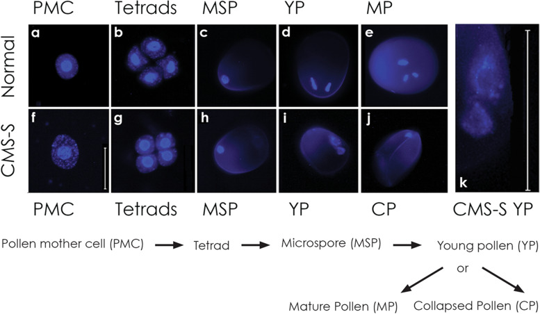
Background: Cytoplasmic male sterility (CMS) is a maternally inherited failure to produce functional pollen that most commonly results from expression of novel, chimeric mitochondrial genes. In Zea mays, cytoplasmic male sterility type S (CMS-S) is characterized by the collapse of immature, bi-cellular pollen. Molecular and cellular features of developing CMS-S and normal (N) cytoplasm pollen were compared to determine the role of mitochondria in these differing developmental fates.

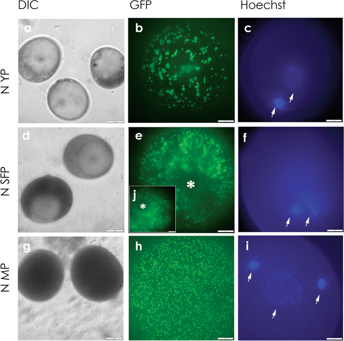
Results: Terminal deoxynucleotidyl transferase dUTP nick end labeling revealed both chromatin and nuclear fragmentation in the collapsed CMS-S pollen, demonstrating a programmed cell death (PCD) event sharing morphological features with mitochondria-signaled apoptosis in animals. Maize plants expressing mitochondria-targeted green fluorescent protein (GFP) demonstrated dynamic changes in mitochondrial morphology and association with actin filaments through the course of N-cytoplasm pollen development, whereas mitochondrial targeting of GFP was lost and actin filaments were disorganized in developing CMS-S pollen. Immunoblotting revealed significant developmental regulation of mitochondrial biogenesis in both CMS-S and N mito-types. Nuclear and mitochondrial genome encoded components of the cytochrome respiratory pathway and ATP synthase were of low abundance at the microspore stage, but microspores accumulated abundant nuclear-encoded alternative oxidase (AOX). Cytochrome pathway and ATP synthase components accumulated whereas AOX levels declined during the maturation of N bi-cellular pollen. Increased abundance of cytochrome pathway components and declining AOX also characterized collapsed CMS-S pollen. The accumulation and robust RNA editing of mitochondrial transcripts implicated translational or post-translational control for the developmentally regulated accumulation of mitochondria-encoded proteins in both mito-types.

Conclusions: CMS-S pollen collapse is a PCD event coincident with developmentally programmed mitochondrial events including the accumulation of mitochondrial respiratory proteins and declining protection against mitochondrial generation of reactive oxygen species.

Keywords: Cytoplasmic male sterility; Maize; Mitochondria; Pollen development; Programmed cell death.


Conflict of interest statement

The authors declare no competing interests.

Figures

Fig. 1
Fig. 1
Normal (N) and CMS-S pollen development visualized with DAPI stain. A flow diagram of pollen development is presented beneath DAPI-stained images of key developmental stages. a-c N-cytoplasm and f–h CMS-S pollen appearing morphologically identical at the a, f pollen mother cell (PMC); b, g tetrad; and c, h microspore (MSP) stages. d, i Young bi-cellular pollen (YP) with one nucleus fragmenting in CMS-S. e N-cytoplasm, mature, tri-cellular pollen (MP). j Collapsed, bi-cellular CMS-S pollen (CP). k Enlarged detail of nuclei from panel i showing marginalized, fragmented chromatin. Bars = 50 µm
Fig. 2
Fig. 2
Terminal dUTP Nick End Labeling (TUNEL) assay and DAPI staining of normal (N) and CMS-S pollen. a-l Tissue sections of developing maize pollen dual stained with a-f TUNEL and g-l DAPI. a, g TUNEL-negative nuclei in N-cytoplasm young pollen (YP). b, h TUNEL-positive staining in the nuclei of CMS-S YP. c, i Fully collapsed CMS-S pollen (CP) showing condensed cytoplasm with punctate, TUNEL-positive bodies. d, j Starch-filling N-cytoplasm mature pollen (MP) showing no TUNEL staining. e, k TUNEL positive staining of MP pre-treated with DNase. f, l Negative control for e, k. Arrows, TUNEL-positive nuclear regions; circles, condensed, TUNEL-positive cytosol; n, Nuclei. Bars = 50 µm
Fig. 3
Fig. 3
Mitochondrial targeting of green fluorescent protein (GFP) and mitochondrial membrane potential in normal (N) cytoplasm maize pollen development. a-l Scanning laser confocal images of a-d young, bi-cellular pollen (YP); e–h starch filling pollen (SFP); and i-l mature pollen (MP). For each developmental stage, multiple images of the same pollen grain are shown. a, e, i Bright field digital interference contrast (DIC). b, f, j Pollen-expressed, mitochondria-targeted GFP. c, g, k Mitochondrial staining with the potentiometric dye JC-1. d, h, l Merged GFP and JC-1 images. Bars = 10 µm
Fig. 4
Fig. 4
Mitochondrial targeting of green fluorescent protein (GFP) and loss of mitochondrial membrane potential in CMS-S maize pollen development. a-l Scanning laser confocal images of a-d young, bi-cellular pollen (YP); e–h pre-collapse, starch-filling pollen (PC); and i-l collapsed pollen (CP). For each developmental stage, multiple images of the same pollen grain are shown. a, e, i Bright field digital interference contrast (DIC). b, f, j Pollen-expressed, mitochondria-targeted GFP. c, g, k Mitochondrial staining with the potentiometric dye JC-1. d, h, l Merged GFP and JC-1 images. Bars = 25 µm for a-d; 10 µm for e-l
Fig. 5
Fig. 5
Mitochondrial morphology changes in normal (N) cytoplasm maize pollen development. a-j Spinning disc confocal micrographs of a-c young, bi-cellular pollen (YP); d-f starch filling pollen (SFP); and g-i mature pollen (MP). For each developmental stage, images of different pollen grains collected from the same anther are shown; a, d, g Bright field digital interference contrast (DIC). b, e, h, j Mitochondria-targeted green fluorescent protein (GFP). c, f, i Hoechst nuclear staining. j inset showing a transition stage between e and h. *, germination pore; arrows, Hoechst-stained nuclei. Bars = 20 µm for a, d, g; 5 µm for b, c, e, f, h, i, j
Fig. 6
Fig. 6
Actin filament organization and co-localization of mitochondria and actin filaments in maize pollen development. Spinning disc confocal micrographs of Phalloidin-iFluor 532 conjugate stained a-c normal (N) cytoplasm and d-e CMS-S pollen. a, d Lose actin network at the young pollen (YP) stage. b, c Organized, parallel actin arrays in N-cytoplasm starch-filling pollen (SFP) and mature pollen (MP), respectively. Parallel filaments run from the germination pore to the opposite side of the MP grain. e Disorganized, short actin filaments in CMS-S collapsed pollen (CP). f–h Spinning disc confocal micrographs of N-cytoplasm SFP. f Mitochondria-targeted green fluorescent protein (GFP) and g Phalloidin-iFluor 532 conjugate stained actin filaments (f-actin) of the same pollen grain. h Merged f and g images showing co-alignment of mitochondria with actin filaments. *, germination pore. Bars = 5 µm
Fig. 7
Fig. 7
Developmentally regulated mitochondrial biogenesis in maize microspores and pollen. a Immunodetection of mitochondrial proteins in developing maize microspores and pollen. Proteins (ATP1, ATP2, ATP 6, ATP8, mitochondrial ATP synthase subunits 1, 2, 6 and 8, respectively; COXII, cytochrome oxidase subunit 2; NAD7, NADH dehydrogenase subunit 7; AOX, alternative oxidase) and the PORIN loading control were immunodetected following denaturing gel electrophoresis. Cropped blot images are shown boxed, and full length blots are presented in Additional File 2, Supplementary Fig. 2. Protein samples contained: Lane 1, 1 ug of protein extracted from normal (N) cytoplasm immature ear mitochondrial pellets; lanes 2–7, 10 ug of total protein extracted from N or CMS-S (S) microspore (MSP), or pollen stages: collapsed pollen (CP), young pollen (YP), starch filling pollen (SFP), mature pollen (MP). b-e Accumulation of mitochondrial proteins from CMS-S and N-cytoplasm maize microspores and developing pollen as compared to mature, N-cytoplasm pollen (set equal to 1). Error bars correspond to the standard deviation for three biological replicates. q,r,s different letters distinguish samples that differ in the relative abundance of ATP1, COXII, ATP6 or AOX at p ≤ 0.05 by a post-ANOVA Tukey’s HSD test. x,y,z different letters distinguish samples that differ in the relative abundance of ATP2, ATP8 or NAD7 at p ≤ 0.05 by the same test

Similar articles

Cited by

References

    1. Hanson MR, Bentolila S. Interactions of mitochondrial and nuclear genes that affect male gametophyte development. Plant Cell. 2004;16(Suppl):S154–69. - PMC - PubMed
    1. Zubko MK. Mitochondrial tuning fork in nuclear homeotic functions. Trends Plant Sci. 2004;9:61–64. doi: 10.1016/j.tplants.2003.12.001. - DOI - PubMed
    1. Linke B, Borner T. Mitochondrial effects on flower and pollen development. Mitochondrion. 2005;5:389–402. doi: 10.1016/j.mito.2005.10.001. - DOI - PubMed
    1. Carlsson J, Leino M. Sohlberg J Sundstrom JF, Glimelius K. Mitochondrial regulation of flower development Mitochondrion. 2008;8:74–86. - PubMed
    1. Horn R, Gupta KJ, Colombo N. Mitochondrion role in molecular basis of cytoplasmic male sterility. Mitochondrion. 2014;19:198–205. doi: 10.1016/j.mito.2014.04.004. - DOI - PubMed

LinkOut - more resources